This guide is general education for pet owners. If your pet has urgent symptoms, possible poisoning, injury, breathing trouble, collapse, severe pain, or sudden decline, contact a licensed veterinarian or local emergency service.
Editorial standard
How this guide was prepared
Acute glaucoma represents the supreme ocular emergency in veterinary medicine, characterized by an acute, pathological surge in intraocular pressure (IOP) that far exceeds the physiological tolerance of the retinal ganglion cells and optic nerve axons. In healthy dogs, continuous production of aqueous humor by the ciliary body is balanced by fluid outflow through the drainage system at the iridocorneal angle (the filtration angle and trabecular meshwork), maintaining normal intraocular pressure between 15 and 25 mmHg. In acute glaucoma—most frequently primary angle-closure glaucoma (PACG) secondary to hereditary pectinate ligament dysplasia, or secondary to anterior uveitis or lens luxation—the drainage pathway suddenly collapses or becomes obstructed. Intraocular pressure surges dramatically to 40, 60, or even 80 mmHg. Within 24 to 48 hours of sustained ocular hypertension, retinal ganglion cells suffer irreversible ischemic death, and optic nerve axonal transport ceases permanently. What begins as a red, squinting eye in the morning can result in permanent, untreatable blindness by the following afternoon unless emergency medical decompression is achieved.
Quick Answer: Recognizing and Acting on Acute Dog Glaucoma
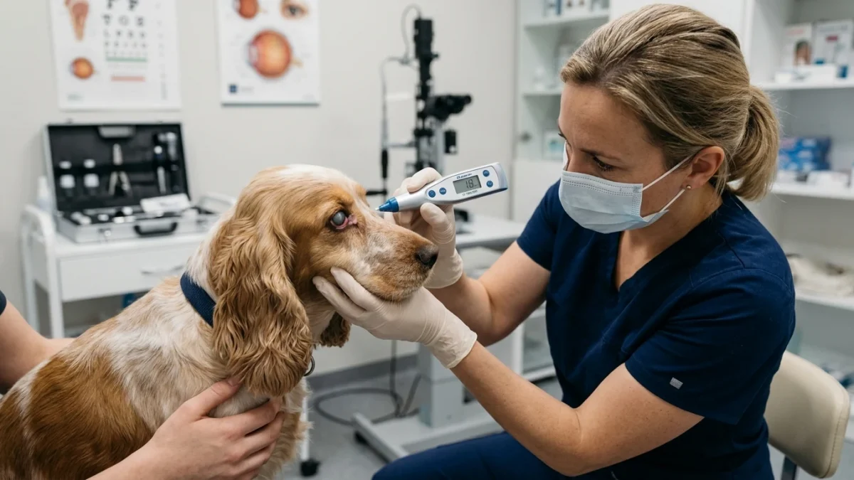
If your dog suddenly develops a cloudy, hazy blue-gray cornea, intense redness in the white of the eye (engorged episcleral vessels), severe squinting (blepharospasm), and a pupil that remains dilated or sluggishly mid-dilated in bright light, suspect acute glaucoma. This condition is excruciatingly painful, causing pets to become head-shy, depressed, and refuse food. Do not wait for a regular daytime appointment. Transport your dog immediately to an emergency facility. Emergency clinicians perform rebound or applanation tonometry (using a TonoVet or Tono-Pen) to measure eye pressure. Pressures above 30 mmHg demand immediate topical pressure-lowering medications (carbonic anhydrase inhibitors and prostaglandin analogs) or systemic osmotic diuretics to preserve permanent vision.
Pathophysiology: How High Pressure Destroys the Canine Retina
The internal architecture of the canine eye is exquisitely delicate, and elevated intraocular pressure inflicts multi-tissue destruction through distinct pathological mechanisms:
1. Retinal Ganglion Cell Ischemia: When intraocular pressure rises above 30 to 40 mmHg, perfusion pressure in the delicate retinal capillaries collapses. Oxygen and glucose supply to the photoreceptors and retinal ganglion cells ceases. Cellular apoptosis begins within hours; complete and irreversible loss of retinal electrical activity occurs after 24 to 48 hours of sustained pressure.
2. Optic Nerve Axonal Crushing (Optic Cupping): High pressure exerts physical force against the cribriform plate at the posterior pole of the globe, interrupting retrograde axonal transport of vital neurotrophic factors from the brain. The optic nerve head becomes deeply excavated or “cupped,” producing permanent optic atrophy.
3. Corneal Edema: The corneal endothelial monolayer maintains corneal clarity by actively pumping fluid out of the corneal stroma. When IOP surges, the hydrostatic pressure forces aqueous humor directly into the stroma, overwhelming endothelial pumps and turning the normally crystal-clear cornea into a hazy, milky blue-gray glass.
4. Episcleral Vascular Congestion (“Medusa Head”): Because aqueous humor cannot drain through internal pathways, venous blood backs up into external scleral vessels. The white of the eye becomes crisscrossed with thick, deep red, tortuous episcleral vessels that do not blanch with topical phenylephrine (distinguishing glaucoma from superficial conjunctivitis).
Clinical Emergency Triage Matrix
| Triage Tier | Clinical Presentation | Required Action |
|---|---|---|
| Tier 1: Acute Ocular Hypertension & Threat of Blindness (0-12h) | Acute onset (<12 hours) of intense squinting, cloudiness (blue-gray cornea), fixed mid-dilated pupil; prominent ropy red scleral vessels; vision is still present (positive menace response or dazzle reflex); pet is head-shy and whimpering. | Transport immediately to an emergency veterinary hospital with 24-hour tonometry. Immediate medical reduction of IOP within this 12-hour golden window has a high probability of preserving permanent vision. Avoid pressure on the neck (use a harness, not a collar). |
| Tier 2: Established Hypertension with Vision Loss (12-36h) | Cornea is densely opaque; menace response is absent (dog cannot see hands approaching eye); pupil is fixed and unresponsive to light; eye may appear subtly enlarged; pet is depressed and rubbing head against carpet. | Proceed directly to an emergency clinic or veterinary ophthalmology referral center. Aggressive medical decompression can sometimes restore electrical function to stunned retinal cells if intervention occurs within 36 hours. Prompt therapy is also vital to initiate preventative treatment in the unaffected companion eye. |
| Tier 3: Chronic Buphthalmos & End-Stage Blindness (>48h) | Eyeball is visibly stretched and enlarged (buphthalmos); lens is luxated or subluxated; white stretch marks (Haab’s striae) visible on cornea; complete irreversible blindness. | Schedule a comprehensive veterinary ophthalmic evaluation. While vision cannot be regained in end-stage buphthalmic eyes, long-term pain management, ciliary ablation, or gentle surgical enucleation is required to eliminate chronic, severe ocular headache. |
Dangerous Home Pitfalls: What NOT to Do (Rule S1)
- NEVER administer atropine eye drops: Atropine induces profound pupil dilation (mydriasis). In an eye with compromised drainage angles, dilating the pupil folds the iris into the iridocorneal junction, shutting down remaining drainage and driving eye pressure beyond 60 to 80 mmHg.
- NEVER apply corticosteroid eye drops without veterinary fluorescein staining: Steroids suppress corneal healing. If the red, squinting eye is caused by an unnoticed corneal scratch rather than glaucoma, topical steroids provoke rapid collagenolytic “corneal melting,” resulting in globe rupture within days.
- NEVER walk your dog on a neck collar or choke chain: Pulling against a neck collar compresses the jugular veins, increasing intracranial and jugular venous pressure, which immediately transmits to the ophthalmic venous plexus and raises intraocular pressure by 4 to 8 mmHg. Always use a chest harness.
- NEVER assume a red eye is just “pink eye” (conjunctivitis): Benign conjunctivitis involves superficial movable vessels, a clear cornea, a normal reactive pupil, and normal eye pressure. Treating acute glaucoma as simple conjunctivitis is the leading cause of preventable permanent blindness in dogs.
Diagnostic Standards: Rebound Tonometry & Fluorescein Staining
Definitive diagnosis of glaucoma requires an accurate, numerical measurement of intraocular pressure using an electronic rebound tonometer (such as the TonoVet) or applanation tonometer (Tono-Pen):
- Baseline Tonometry: The tonometer gently taps the central cornea multiple times to calculate mean pressure. Normal canine IOP is 15 to 25 mmHg. A pressure reading above 25 to 30 mmHg in an inflamed eye confirms ocular hypertension, while readings of 40 to 80 mmHg signify acute fulminant glaucoma.
- Bilateral Assessment: Both eyes must always be measured. In primary glaucoma, while one eye presents in acute crisis, the fellow eye frequently possesses the identical anatomical drainage defect (goniodysgenesis) and requires immediate prophylactic medical therapy.
- Fluorescein Stain Testing: Prior to instilling any topical ocular medication, a fluorescein dye strip is applied to verify corneal epithelial integrity. An intact green glow indicates a corneal ulcer, altering pharmacological choices.
Emergency Medical Stabilization and Vision Preservation
The immediate medical objective is rapid, aggressive pressure reduction to restore retinal perfusion before permanent ischemic necrosis occurs:
1. Topical Prostaglandin Analogs: Ophthalmic prostaglandin F2-alpha analogs (such as latanoprost) are the most potent medical agents for acute canine primary glaucoma. Latanoprost induces intense miosis (pupil constriction), pulling the iris root away from the drainage angle and dramatically increasing unconventional uveoscleral outflow, often dropping pressure from 60 mmHg down to normal levels within 45 to 60 minutes. (Note: latanoprost is contraindicated if anterior lens luxation is present).
2. Topical Carbonic Anhydrase Inhibitors (CAIs): Medications such as dorzolamide or brinzolamide inhibit the enzyme carbonic anhydrase in the ciliary body epithelium, reducing aqueous humor production by 40% independent of pupil size or uveoscleral pathways.
3. Systemic Osmotic Diuretics (Mannitol): In refractory cases where pressures remain dangerously elevated above 50 mmHg despite topical therapy, intravenous mannitol is administered over 20 to 30 minutes. Mannitol increases intravascular blood osmolarity, drawing water out of the vitreous humor and collapsing vitreous volume, rapidly decompressing the eye.
4. Fellow Eye Prophylaxis: In primary glaucoma, the unaffected eye is immediately started on preventative topical therapy (such as daily dorzolamide or betaxolol) to significantly delay the onset of glaucoma in the companion eye.
Frequently Asked Questions
Can a dog feel pain from acute glaucoma?
Yes, acute glaucoma is agonizingly painful, often described as an excruciating migraine or intense ocular pressure that radiates through the entire skull. However, dogs rarely cry or whimper; instead, they display subtle pain behaviors: becoming withdrawn, hiding under beds, sleeping constantly, refusing food, flinching when petted near the head (head-shyness), or aggressively rubbing the affected eye against carpets and paws.
Which dog breeds are most prone to acute primary glaucoma?
Primary angle-closure glaucoma has strong genetic links in Cocker Spaniels (both American and English), Basset Hounds, Chow Chows, Siberian Huskies, Shar-Peis, Boston Terriers, and Beagles. In these breeds, hereditary malformation of the drainage angle (pectinate ligament dysplasia) creates a lifelong risk of sudden, bilateral angle closure.
What if medical eye drops fail to keep the eye pressure down?
If medical therapy cannot maintain intraocular pressure in a safe range, surgical interventions by a board-certified veterinary ophthalmologist are considered. Options include endocyclophotocoagulation (ECPC laser ablation of fluid-producing ciliary processes), drainage shunt placement (Ahmed valve), or, if permanent blindness and irreversible pain have developed, surgical enucleation (eye removal) or intrascleral prosthesis to ensure permanent, pain-free comfort.
Scientific Sources and Literature
- Miller PE, Bentley E. “Clinical signs and diagnosis of canine glaucoma: the role of rebound tonometry and gonioscopy.” Veterinary Clinics of North America: Small Animal Practice. PMID: 29959652
- Kato K, et al. “Incidence and risk factors of primary angle-closure glaucoma in purebred canines: a multi-center study.” Journal of the American Veterinary Medical Association. PMID: 31086219
- Gould D, et al. “Medical and surgical management of acute primary glaucoma in dogs: vision retention and long-term outcomes.” Veterinary Ophthalmology. PMID: 24397805
Caregiver Emergency Glaucoma Action Checklist
- Inspect the eye closely: note if the cornea is cloudy (blue-gray) and if the white of the eye is deep crimson red.
- Check pupil symmetry: observe whether the affected pupil is larger than the normal pupil and fails to shrink in light.
- Test vision gently: observe if your dog blinks when your hand makes a threatening gesture toward the eye (menace test).
- NEVER administer leftover human or pet eye drops containing atropine or steroids.
- NEVER walk the dog on a neck collar; switch immediately to a loose body harness to prevent jugular compression.
- Transport your pet immediately to an emergency veterinary hospital equipped with an electronic tonometer.
Source transparency
Reference mix for this guide
Source labels describe the type of organization behind each reference; they are not a claim that any outside organization reviewed this article.
- Dog Red Eye & Squinting: Glaucoma vs Conjunctivitis Reference source · newspet.net
- Dog Eye Proptosis & Ocular Trauma Emergency Reference source · newspet.net
- Dog Shaking & Lethargic Differential Triage Reference source · newspet.net
- Dog First Seizure & Neurological Emergency Guide Reference source · newspet.net
- PMID: 29959652 Government or public agency · pubmed.ncbi.nlm.nih.gov
- PMID: 31086219 Government or public agency · pubmed.ncbi.nlm.nih.gov
- PMID: 24397805 Government or public agency · pubmed.ncbi.nlm.nih.gov