This guide is general education for pet owners. If your pet has urgent symptoms, possible poisoning, injury, breathing trouble, collapse, severe pain, or sudden decline, contact a licensed veterinarian or local emergency service.
Editorial standard
How this guide was prepared
Traumatic ocular proptosis—the sudden anterior displacement of the eyeball beyond the orbital rim, with the eyelids becoming entrapped behind the equator of the globe—is one of the most acute, visually distressing surgical emergencies in veterinary medicine. Because the globe is physically trapped outside the orbit, venous return is obstructed while arterial supply continues, resulting in massive retrobulbar congestion, extreme intraocular pressure elevation, and progressive corneal desiccation. Furthermore, forward traction on the optic nerve and avulsion of delicate extraocular muscles (particularly the medial rectus) threaten permanent, irreversible blindness within hours. Immediate emergency stabilization and urgent surgical reduction are imperative to preserve vision and salvage the globe.
Quick Answer: First Aid for a Dislocated Eyeball
If your dog’s eye has popped out of its socket, keep the dog calm and immediately protect the exposed cornea from drying out. Generously apply sterile artificial tears, ophthalmic lubricant, or saline-soaked sterile gauze over the entire eye. Do NOT attempt to push the eyeball back in. Apply an Elizabethan collar or gently restrain your dog’s paws to prevent catastrophic self-trauma. Transport the dog to an emergency veterinary surgical facility immediately; surgical reduction within the first 1 to 2 hours provides the highest probability of saving the eye.
Breed Predispositions: Why Brachycephalic Dogs Are at Extreme Risk
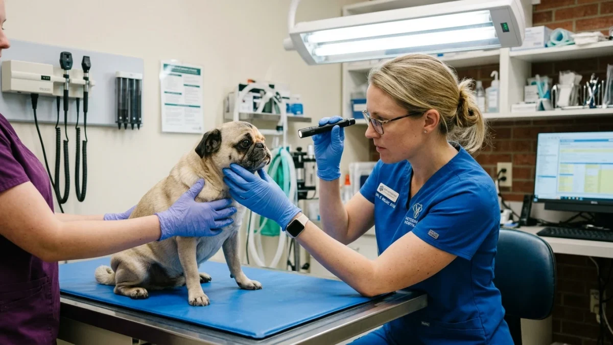
Canine ocular anatomy varies drastically across skull conformations. Brachycephalic breeds—including Pugs, French Bulldogs, Boston Terriers, Shih Tzus, and Pekingese—possess shallow, flattened bony orbits paired with excessively wide eyelid openings (macroblepharon or exophthalmos). In these breeds, the eyeball sits precariously forward with minimal bony protection. Consequently, minor blunt trauma—such as vigorous play with another dog, accidental head bumping against furniture, or even excessive scruff restraint during grooming—can cause the globe to pop forward past the eyelids.
In contrast, mesocephalic and dolichocephalic dogs (such as German Shepherds or Collies) possess deep bony orbital sockets that securely encase the eyeball. In deep-socketed breeds, ocular proptosis occurs only as a result of severe, massive head trauma, such as vehicular accidents or dog attack bite crush injuries. In these non-brachycephalic patients, proptosis is frequently accompanied by concurrent skull fractures, brain contusions, and systemic shock.
Clinical Emergency Triage Matrix
| Triage Tier | Clinical Presentation | Required Action |
|---|---|---|
| Tier 1: Immediate Emergency (0-2h) | Eyeball visibly displaced outside the orbital rim with eyelids trapped behind the globe; globe appears congested, dark red, or hazy; dog vocalizing in agony; bleeding from the orbit; concurrent head trauma, epistaxis (nosebleeds), or loss of consciousness. | Apply generous sterile eye lubrication or moist sterile saline gauze. Keep dog immobilized. Transport immediately to an emergency veterinary surgical center for emergency general anesthesia and temporary tarsorrhaphy. |
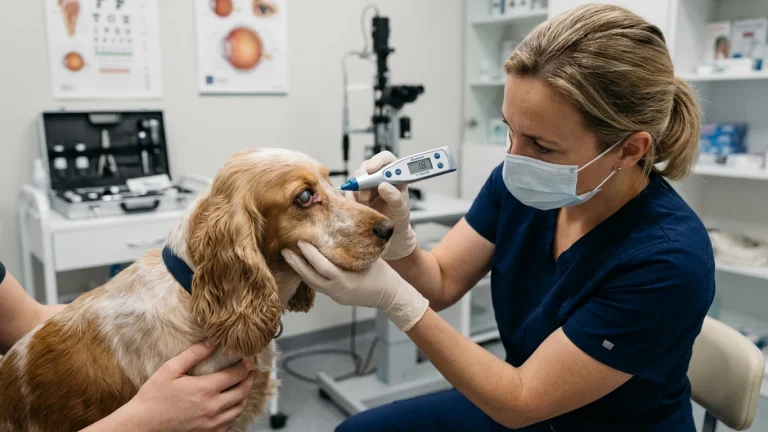
| Tier 2: Urgent Ophthalmic Evaluation (2-6h) | Eyeball partially protruding with severe eyelid swelling (chemosis) and intense blepharospasm (tight squinting), but eyelids are still able to close partially over the cornea; severe corneal laceration or deep foreign object penetration. | Apply sterile eye lubricant and an Elizabethan collar immediately. Proceed directly to a veterinary ophthalmology specialist or 24-hour emergency hospital for detailed slit-lamp and fluorescein diagnostics. |
| Tier 3: Same-Day Ocular Care (6-12h) | Mild ocular trauma with clear tearing, normal globe positioning, intact pupil responsiveness, and absence of visible corneal scratches or cloudiness. | Prevent eye rubbing with an Elizabethan collar. Schedule an urgent outpatient veterinary visit for fluorescein stain evaluation to rule out microscopic corneal ulcerations. |
Dangerous Home Pitfalls: What NOT to Do (Rule S1)
- NEVER attempt to push the eyeball back into the socket at home: Attempting home reduction without general anesthesia, lateral canthotomy, and specialized ophthalmic instruments causes catastrophic corneal rupture, vitreous extrusion, and permanent optic nerve transection.
- NEVER apply dry gauze, tissues, or paper towels to the eye: Dry paper and textiles adhere to the fragile corneal epithelium. When pulled away, they strip the epithelial layer, transforming a manageable proptosis into a catastrophic melting corneal ulcer.
- NEVER use medicated human eye drops (Visine, steroid drops): Vasoconstrictor drops (Visine) worsen ocular tissue ischemia. Steroid eye drops on a compromised or scratched cornea trigger rapid bacterial colonization and stromal collagen melting. Use ONLY non-medicated sterile saline or pure artificial tear lubricant.
- NEVER leave the dog unattended without physical paw restraint: In agonizing ocular pain, a dog will instinctively paw violently at the displaced eye. A single swipe can shred the cornea or tear the globe completely from the remaining rectus muscles.
Pathology of Ocular Damage During Proptosis
When the globe displaces anterior to the eyelid margins, three destructive biological processes begin simultaneously:
Corneal Desiccation and Exposure Keratitis: The cornea relies on continuous blinking to distribute the precorneal tear film, which provides oxygen and nutrients to the avascular corneal epithelium. With the eyelids trapped behind the globe, the eye cannot blink. Within 15 to 30 minutes of open air exposure, the corneal epithelium dries, cracks, and ulcerates, allowing aggressive bacterial pathogens (such as Pseudomonas) to invade the stroma.
Optic Nerve Traction and Ischemia: The optic nerve and retinal blood vessels enter the posterior pole of the globe. As the eye pops forward, extreme mechanical tension stretches the optic nerve. If stretched beyond its elastic threshold, axonal fibers shear, and central retinal arterial blood flow is severed. If ischemia persists for more than 1 to 2 hours, permanent amaurosis (complete blindness) occurs, even if the eyeball itself is later successfully replaced in the socket.
Extraocular Muscle Avulsion: The globe is anchored by seven extraocular muscles. During traumatic forward displacement, the shortest and most vulnerable muscle—the medial rectus—frequently tears away from the sclera. Avulsion of the medial rectus produces a permanent lateral strabismus (outward deviation of the eye) post-recovery.
Frequently Asked Questions
Can a dog still see after their eye pops out?
Vision retention depends heavily on skull conformation and the speed of surgical reduction. In brachycephalic dogs with minimal trauma reduced within 1 to 2 hours, approximately 20 to 30 percent regain partial or full functional vision. In deep-chested, non-brachycephalic dogs where proptosis required severe blunt force, the visual prognosis is poor (under 5 percent), but surgical replacement is still frequently performed to salvage the cosmetic appearance of the eye.
What is a temporary tarsorrhaphy?
A temporary tarsorrhaphy is the definitive surgical procedure used to manage proptosis. Under general anesthesia, the surgeon performs a small lateral cut (canthotomy) to relieve eyelid tension, gently maneuvers the lubricated globe back into the orbit, and sutures the upper and lower eyelids shut over protective stents. The eyelids remain closed for 14 to 21 days, acting as a natural, vascularized biological bandage while extraocular tissues heal.
When does a proptosed eye have to be removed (enucleated)?
A veterinarian must perform enucleation (surgical removal of the eye) if the globe is physically ruptured, if the cornea has undergone full-thickness necrosis, if more than two or three extraocular muscles are completely avulsed, or if active intraocular hemorrhage (hyphema) indicates complete structural collapse. Enucleation eliminates chronic pain and prevents ascending orbital infection.
Scientific Sources and Literature
- Gilger BC, et al. “Traumatic ocular proptosis in dogs and cats: 84 cases (1980-1993).” Journal of the American Veterinary Medical Association. PMID: 24792442
- Maggs DJ, et al. “Slatter’s Fundamentals of Veterinary Ophthalmology.” Veterinary Ophthalmology. PMID: 25176161
- Gelatt KN. “Veterinary Ophthalmic Surgery and Emergency Globe Repositioning.” Veterinary Surgery. PMID: 28844883
Caregiver Emergency Eye Proptosis Checklist
- Stay calm; keep your dog immobilized and prevent rapid running or shaking of the head.
- Generously soak the protruding eye with sterile saline solution or non-medicated artificial tear ophthalmic lubricant.
- Cover the eye loosely with a clean, saline-moistened gauze pad; never apply dry cloths or paper towels.
- Fit an Elizabethan collar immediately, or gently hold the dog’s front legs to prevent paw scratching.
- Never attempt to manipulate the eye or press it back into the socket.
- Proceed immediately to the nearest open emergency veterinary hospital with surgical capabilities.
Source transparency
Reference mix for this guide
Source labels describe the type of organization behind each reference; they are not a claim that any outside organization reviewed this article.
- Dog Red Eye & Glaucoma Differential Triage Reference source · newspet.net
- Dog Bee Sting & Facial Swelling Emergency Reference source · newspet.net
- Dog Bleeding Wound & Hemorrhage First Aid Reference source · newspet.net
- Dog Shaking & Lethargic Differential Triage Reference source · newspet.net
- PMID: 24792442 Government or public agency · pubmed.ncbi.nlm.nih.gov
- PMID: 25176161 Government or public agency · pubmed.ncbi.nlm.nih.gov
- PMID: 28844883 Government or public agency · pubmed.ncbi.nlm.nih.gov