This guide is general education for pet owners. If your pet has urgent symptoms, possible poisoning, injury, breathing trouble, collapse, severe pain, or sudden decline, contact a licensed veterinarian or local emergency service.
Dosages must be calculated by a licensed veterinarian based on species, weight, and bloodwork. Never administer human medications or self-adjust prescription doses for pets.
Editorial standard
How this guide was prepared
Post-operative recovery requires strict adherence to discharge instructions to prevent surgical failure, dehiscence, or secondary infection. The first 48 to 72 hours demand intensive observation, environmental restriction, and precise pharmacological management.
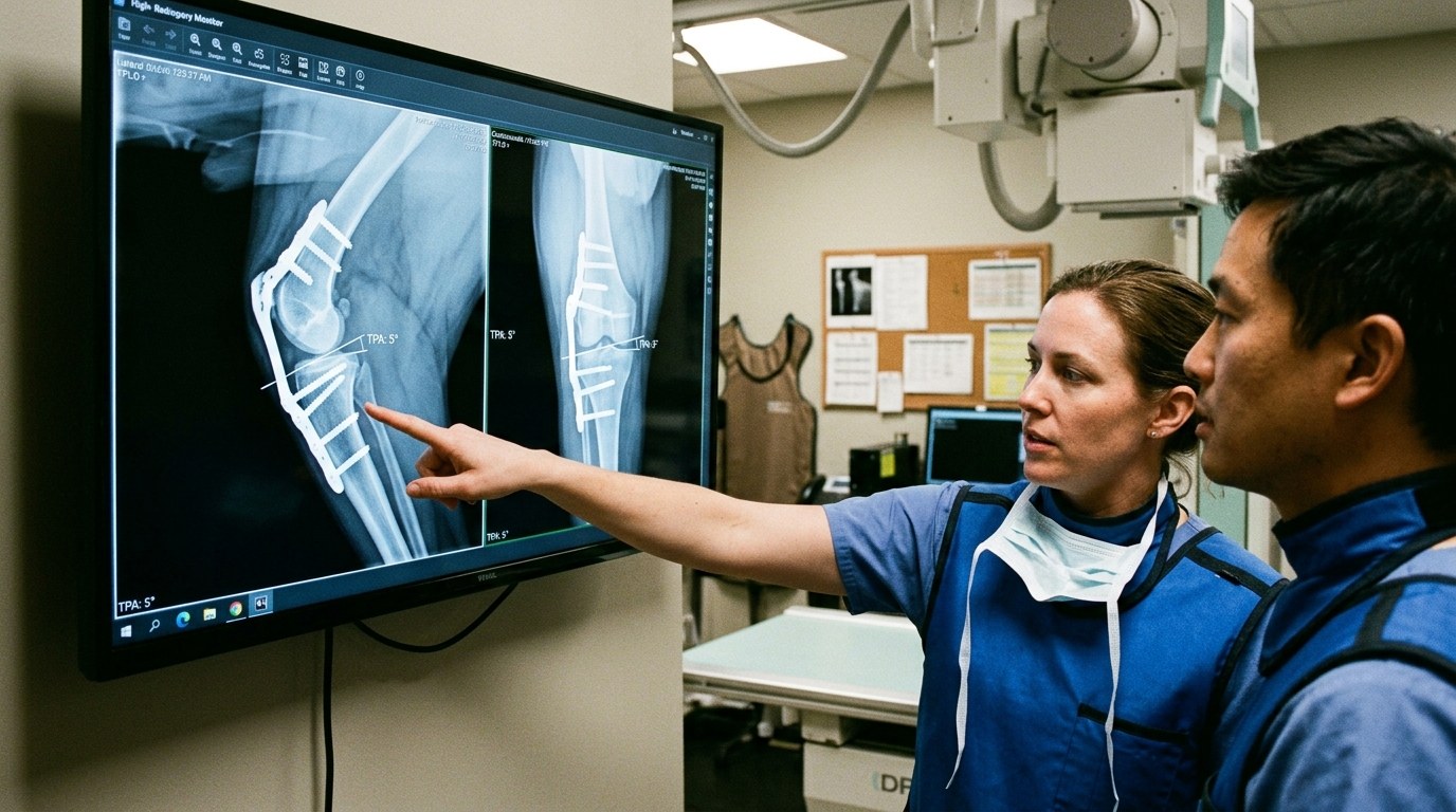
Strict 8-Week Confinement Mandate: Tibial Plateau Leveling Osteotomy (TPLO) involves creating a full semicircular radial cut through the proximal tibia, rotating the plateau to neutralize cranial tibial thrust, and securing the bone fragments with a rigid titanium locking plate and cortical screws. The surgical plate is a temporary holding bridge—it CANNOT withstand the mechanical forces of jumping, running, or playing before complete biological bone union (typically 8 weeks). Uncontrolled activity causes screw pull-out, catastrophic tibial fracture, or meniscal tears requiring revision surgery.
Clinical Quick Answer & Immediate Actions
Surgical success relies on strict postoperative confinement, multimodal analgesia, and vigilant incision monitoring. Unsupervised access to incisions compromises the surgical closure. Caregivers must restrict activity entirely and administer all medications strictly as prescribed.
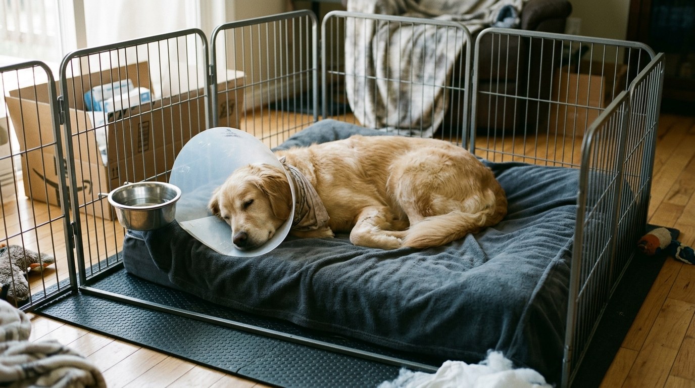
Incision sites must be inspected twice daily for erythema, purulent discharge, or unexpected swelling. A properly fitted Elizabethan collar (E-collar) or recovery suit is non-negotiable; patients can destroy surgical closures within seconds of unsupervised access.
Pharmacology and Pain Management
Analgesic protocols typically utilize a multimodal approach, combining non-steroidal anti-inflammatory drugs (NSAIDs) with opioids or gabapentinoids. This strategy targets different pain pathways while minimizing adverse dose-dependent effects.
Administer all prescribed antibiotics until the entire course is completed to prevent antimicrobial resistance, even if the surgical site appears fully healed. Never substitute veterinary prescriptions with human over-the-counter pain relievers, which are highly toxic to companion animals.
| Post-Op Day | Clinical Focus | Expected Finding |
|---|---|---|
| Days 1-3 | Pain control & strict rest | Mild bruising, lethargy, decreased appetite |
| Days 4-7 | Tissue granulation | Decreased swelling, returning appetite |
| Days 10-14 | Suture removal / Recheck | Closed margins, resolution of bruising |
Environmental management is equally critical. Isolate the recovering patient from other household animals. Hardwood floors should be covered with non-slip rugs to prevent orthopedic trauma, and access to stairs or furniture must be blocked entirely.
Dietary reintroduction should begin with small, highly digestible meals (e.g., boiled chicken and rice or veterinary gastrointestinal diets) to accommodate anesthesia-induced nausea and gastrointestinal stasis.
When to Seek Emergency Intervention
Immediate veterinary reassessment is required if the patient exhibits active surgical site bleeding, complete anorexia lasting longer than 36 hours, uncontrolled vomiting, or sudden vocalization indicating breakthrough pain.
Orthopedic and neurological patients presenting with sudden loss of motor function, or those unable to urinate within 24 hours of discharge, constitute a critical emergency requiring immediate clinic transport.
Post-Operative Tracker
Maintain a strict medication log to prevent accidental overdoses. Access our Surgical Recovery Tracker to record dosage times, feeding amounts, and incision observations.
Biomechanical Principles of Cranial Tibial Thrust Neutralization
The canine stifle differs fundamentally from the human knee due to its sloping tibial plateau. In the cranial cruciate ligament (CCL) deficient stifle, weight-bearing generates a forward shearing force (cranial tibial thrust) that forces the femur to slide backward off the tibia:
- Radial Osteotomy & Angle Rotation: Rather than replacing the torn ligament, TPLO surgically alters joint geometry. The surgeon rotates the proximal tibial plateau from an average slope of 28° down to a functional 5° to 6.5°. At this modified angle, gravitational weight-bearing forces are converted into pure axial compression, actively eliminating cranial tibial thrust and stabilizing the stifle joint without requiring an intact cruciate ligament.
- Meniscal Release vs. Late Meniscal Tears: The medial meniscus frequently suffers crush injury during acute CCL tears. Surgeons inspect, debride, and often perform a partial medial meniscectomy or meniscal release. However, in roughly 5% to 8% of dogs, a “late meniscal tear” occurs weeks to months after surgery due to minor rotational micro-instability, presenting as sudden acute non-weight-bearing lameness that requires arthroscopic evaluation.
- Bone Union Stages & 8-Week Orthogonal Radiographs: Bone healing occurs via primary cortical union. Strict confinement is non-negotiable until orthogonal (craniocaudal and lateral) stifle radiographs at 8 weeks confirm bridging bone trabeculae across the osteotomy site. Until bridging callus is verified, implant failure remains a constant risk under uncontrolled loading.
Structured 8-Week Milestone Rehabilitation Protocol
Caregivers must follow a phased, disciplined rehabilitation progression:
- Weeks 1–2 (Acute Inflammatory & Incisional Phase): Absolute crate or exercise-pen confinement on non-slip rugs. Cold pack the stifle for 15 minutes three times daily for the first 5 days. Gentle passive range of motion (PROM) within comfortable limits (10 flexions and extensions twice daily). Elimination breaks on a short 6-foot leash with a supportive belly sling only.
- Weeks 3–4 (Controlled Weight-Bearing Phase): The dog should demonstrate consistent, light toe-touch weight-bearing at a slow walk. Introduce controlled, straight-line leash walks for 5 minutes twice daily on flat grass. Practice weight-shifting exercises by gently nudging the dog’s hips while standing square to encourage equal weight distribution.
- Weeks 5–8 (Strengthening Phase): Gradually advance leash walks by 2 to 3 minutes each week, reaching 15 to 20 minutes of continuous flat walking by week 8. Practice slow sit-to-stand transitions (“dog squats”) to rebuild atrophied quadriceps and biceps femoris muscle mass. Attend the mandatory 8-week orthopedic radiographic recheck for surgical union confirmation.
Frequently Asked Questions
Can I take the E-collar off while I am watching them?
No. Even momentary lapses in supervision can result in the patient accessing the incision. Keep the Elizabethan collar on continuously until the attending veterinarian confirms complete dermal healing (typically 10-14 days).
What is a seroma, and should I be worried?
A seroma is a sterile, fluid-filled pocket that occasionally forms under the incision site due to excess activity. While generally benign, rapid enlargement requires veterinary evaluation to rule out infection or herniation.
My pet hasn’t defecated since surgery. Is this an emergency?
It is common for patients to not defecate for 3 to 5 days post-operatively due to fasting, anesthesia, and opioid analgesics. However, if the patient is straining unproductively, contact the clinic.
Emergency Triage Matrix: Clinical Presentation & Required Response
| Triage Level | Clinical Signs & Presentation | Immediate Action Protocol |
|---|---|---|
| Tier 1: Immediate Emergency (0-2 Hours) | Incision dehiscence (opening), active hemorrhage, pale mucous membranes, extreme lethargy, or respiratory distress. | Wrap the incision site with a clean towel (do not apply tourniquets) and transport immediately to the emergency surgical facility. |
| Tier 2: Urgent Veterinary Care (Same-Day) | Purulent (yellow/green) discharge from incision, localized heat, uncontrolled vomiting, or inability to urinate for >24 hours. | Contact the surgical clinic for a same-day recheck to assess for infection, adjust analgesia, or evaluate renal output. |
| Tier 3: Home Monitoring & Scheduled Care | Mild swelling (seroma), slight redness along the incision margins, or 3-4 days without a bowel movement (if eating normally). | Maintain strict E-collar usage, apply warm compresses only if directed by a veterinarian, and monitor progression at home. |
Surgical Complication Alert
Incision integrity is the most critical factor in postoperative success. Compromises lead to catastrophic failure:
- E-Collar Compliance: Elizabethan collars must remain secured 100% of the time, including during feeding or sleep.
- Activity Restriction: Do not allow running, jumping, or stair climbing until explicitly cleared by the surgical team.
Post-Operative Discharge Checklist
- Verify the exact dosage and frequency of all prescribed analgesics.
- Set up a small, confined recovery area free of elevated surfaces.
- Schedule the 10-to-14-day suture removal or recheck appointment.
Related Clinical Guides & Tools
Peer-Reviewed Clinical References & Evidence Grounding
- Does LLLT improve radiographic healing for dogs with cranial cruciate ligament rupture undergoing TPLO surgery? (opens in new tab) (Veterinary evidence (2025) — Peer-reviewed veterinary study indexed on NCBI PubMed (PMID: 42006389).)
- Canine Cranial Cruciate Ligament Disease (CCLD): A Concise Review of the Recent Literature (opens in new tab) (Animals : an open access journal from MDPI (2025) — Peer-reviewed veterinary study indexed on NCBI PubMed (PMID: 40218423).)
- Associations between meniscal tears and various degrees of osteoarthritis among dogs undergoing TPLO for cranial cruciate ligament rupture (opens in new tab) (BMC research notes (2023) — Peer-reviewed veterinary study indexed on NCBI PubMed (PMID: 36915203).)
- Merck Veterinary Manual: Joint Trauma In Dogs And Cats (opens in new tab) (Verified clinical reference retrieved via Antigravity Ground-Truth Search (merckvetmanual.com).)
Printable tools
Download the matching worksheet
Source transparency
Reference mix for this guide
Source labels describe the type of organization behind each reference; they are not a claim that any outside organization reviewed this article.
- Emergency Triage Protocol Reference source · newspet.net
- Pharmacology Safety Standards Reference source · newspet.net
- Galliprant (Grapiprant) for Dogs: Non-COX Osteoarthritis Triage Reference source · newspet.net
- Trazodone for Dogs: Anxiety, Post-Op Confinement & Triage Reference source · newspet.net
- Post-Operative Pet Rehabilitation: Passive Range of Motion, Cold Laser Therapy, and Hydrotherapy Timelines Reference source · newspet.net
- Dog Mass Removal Aftercare: Seroma Prevention, Histopathology Margins, and Incision Healing Reference source · newspet.net
- Dog Spinal Surgery Recovery (IVDD): Strict Crate Rest Protocols, Bladder Expression, and Neurological Recheck Milestones Reference source · newspet.net
- Cat Limb Amputation Recovery: Pain Management, Litter Box Adaptations, and Tri-Pawd Mobility Reference source · newspet.net