This guide is general education for pet owners. If your pet has urgent symptoms, possible poisoning, injury, breathing trouble, collapse, severe pain, or sudden decline, contact a licensed veterinarian or local emergency service.
Dosages must be calculated by a licensed veterinarian based on species, weight, and bloodwork. Never administer human medications or self-adjust prescription doses for pets.
Editorial standard
How this guide was prepared
Post-operative recovery requires strict adherence to discharge instructions to prevent surgical failure, dehiscence, or secondary infection. The first 48 to 72 hours demand intensive observation, environmental restriction, and precise pharmacological management.
Strict Tension-Relieving Confinement: Curative surgical resection of cutaneous mast cell tumors, soft tissue sarcomas, or infiltrative lipomas requires wide anatomical margins (typically 2 to 3 cm lateral margins and at least one fascial plane deep). Closing these large surgical defects places tremendous tension on cutaneous sutures. Running, jumping, or body scratching will tear deep subcutaneous holding sutures, causing extensive wound dehiscence, hematoma formation, and catastrophic surgical failure. Keep the dog confined on low-traction surfaces with a rigid Elizabethan collar 24/7 for 14 days.
Clinical Quick Answer & Immediate Actions
Surgical success relies on strict postoperative confinement, multimodal analgesia, and vigilant incision monitoring. Unsupervised access to incisions compromises the surgical closure. Caregivers must restrict activity entirely and administer all medications strictly as prescribed.
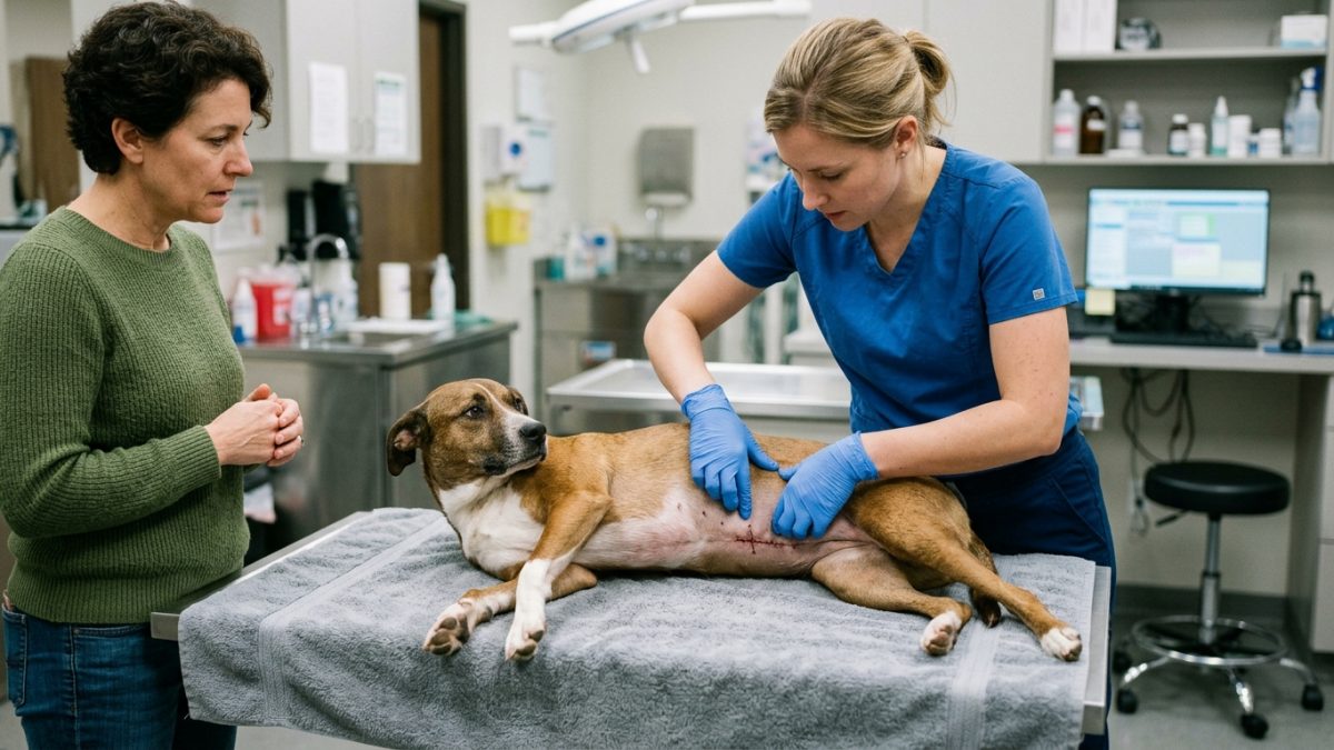
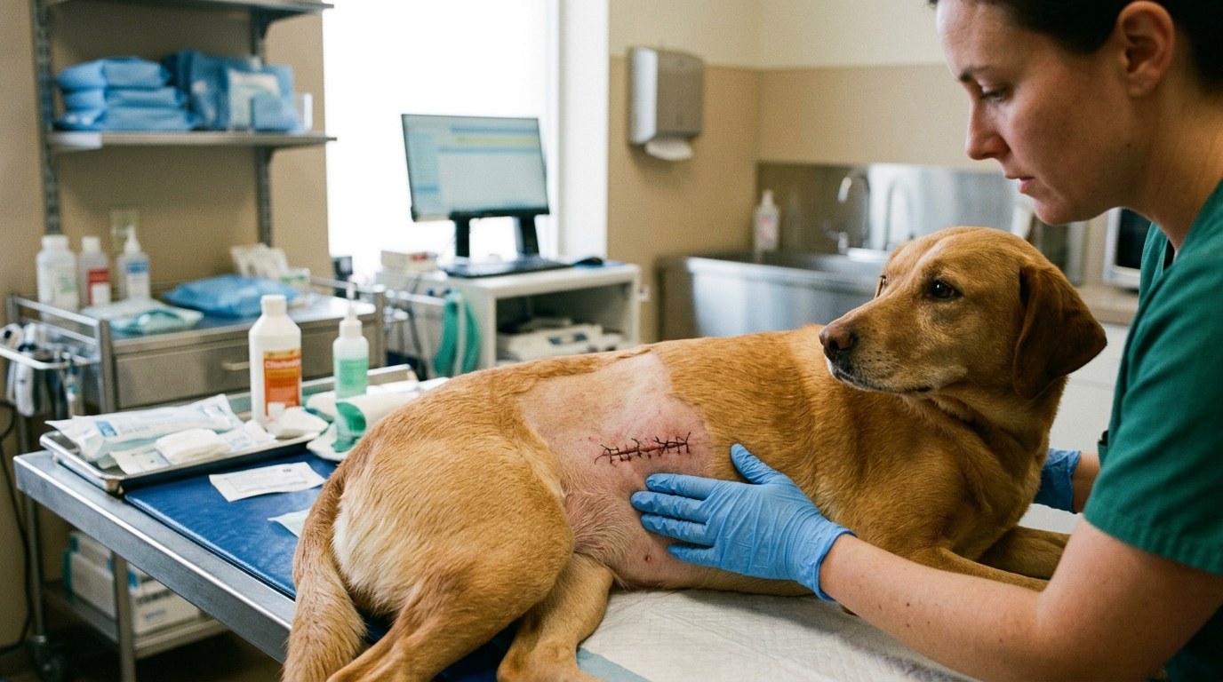
Incision sites must be inspected twice daily for erythema, purulent discharge, or unexpected swelling. A properly fitted Elizabethan collar (E-collar) or recovery suit is non-negotiable; patients can destroy surgical closures within seconds of unsupervised access.
Pharmacology and Pain Management
Analgesic protocols typically utilize a multimodal approach, combining non-steroidal anti-inflammatory drugs (NSAIDs) with opioids or gabapentinoids. This strategy targets different pain pathways while minimizing adverse dose-dependent effects.
Administer all prescribed antibiotics until the entire course is completed to prevent antimicrobial resistance, even if the surgical site appears fully healed. Never substitute veterinary prescriptions with human over-the-counter pain relievers, which are highly toxic to companion animals.
| Post-Op Day | Clinical Focus | Expected Finding |
|---|---|---|
| Days 1-3 | Pain control & strict rest | Mild bruising, lethargy, decreased appetite |
| Days 4-7 | Tissue granulation | Decreased swelling, returning appetite |
| Days 10-14 | Suture removal / Recheck | Closed margins, resolution of bruising |
Environmental management is equally critical. Isolate the recovering patient from other household animals. Hardwood floors should be covered with non-slip rugs to prevent orthopedic trauma, and access to stairs or furniture must be blocked entirely.
Dietary reintroduction should begin with small, highly digestible meals (e.g., boiled chicken and rice or veterinary gastrointestinal diets) to accommodate anesthesia-induced nausea and gastrointestinal stasis.
When to Seek Emergency Intervention
Immediate veterinary reassessment is required if the patient exhibits active surgical site bleeding, complete anorexia lasting longer than 36 hours, uncontrolled vomiting, or sudden vocalization indicating breakthrough pain.
Orthopedic and neurological patients presenting with sudden loss of motor function, or those unable to urinate within 24 hours of discharge, constitute a critical emergency requiring immediate clinic transport.
Post-Operative Tracker
Maintain a strict medication log to prevent accidental overdoses. Access our Surgical Recovery Tracker to record dosage times, feeding amounts, and incision observations.
Oncological Surgical Margins & Histopathological Staging
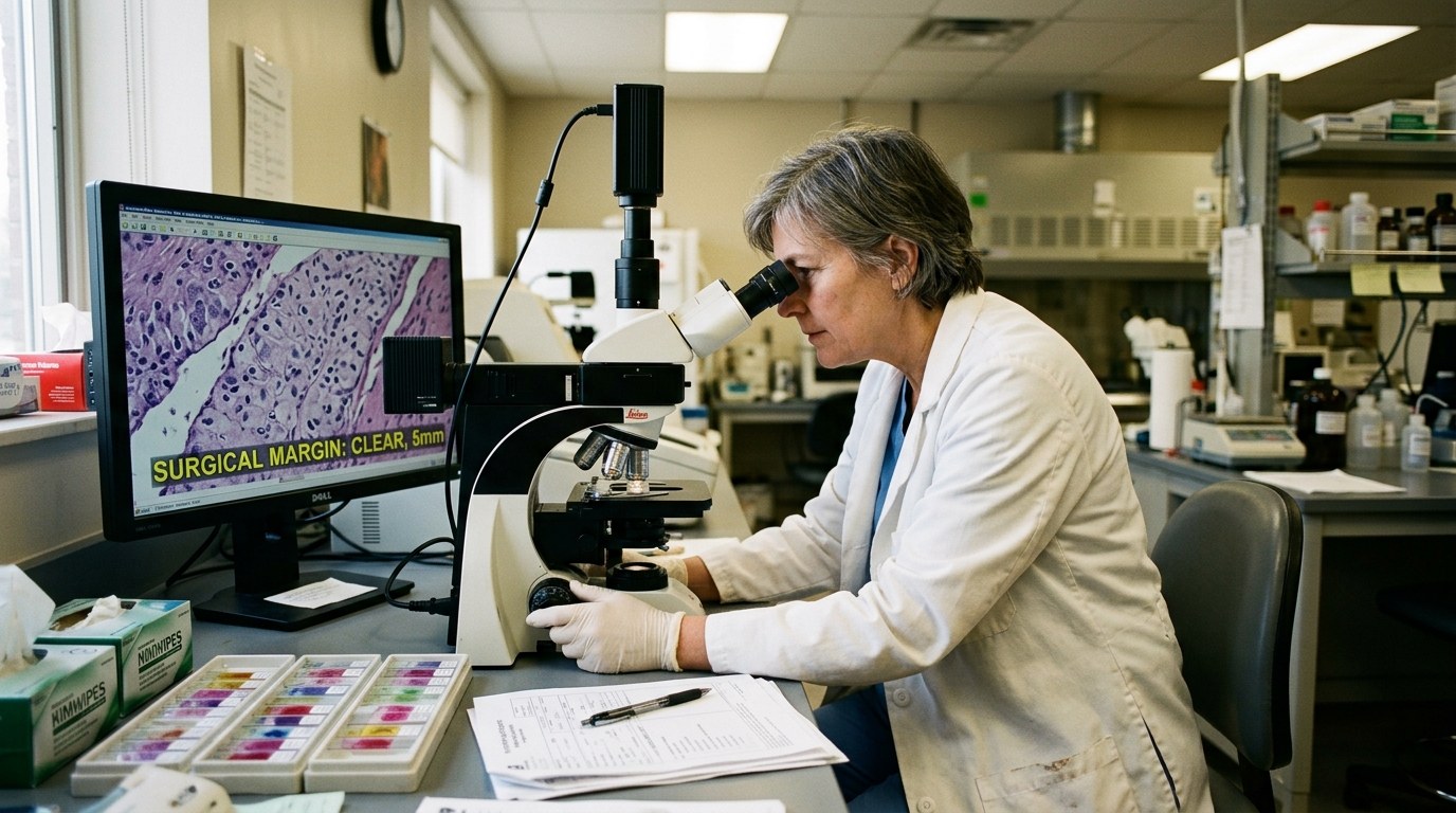
In veterinary surgical oncology, the primary surgical excision represents the single best opportunity for permanent curative disease control. Understanding the pathology report empowers caregivers to manage subsequent clinical steps:
- Clean vs. Incomplete Surgical Margins: The excised tissue specimen is marked with surgical ink and submitted for comprehensive histopathological examination. “Clean” or “negative” margins indicate that no neoplastic cells are visible within 2 to 3 mm of the inked edges. “Incomplete” or “narrow” margins mean cancer cells extend to the surgical border, requiring surgical scar revision, localized radiation therapy, or systemic chemotherapy to prevent local tumor recurrence.
- Managing Active Closed-Suction Drains (Jackson-Pratt): When large skin flaps are mobilized, surgeons frequently place active silicone bulb drains (JP drains) to evacuate inflammatory fluid and collapse dead space. Caregivers must empty the bulb every 8 to 12 hours, record the fluid volume in milliliters, and note fluid character (serosanguinous vs. cloudy purulent). Drains are typically removed once fluid production drops below 2 to 3 milliliters per kilogram per 24-hour period.
- Seroma Formation vs. Wound Infection: Dead space fluid accumulation (seroma) frequently forms beneath large excision sites over the lateral thorax or flank. Seromas appear as soft, cool, painless, fluctuant fluid pockets. Attempting to aspirate a seroma with a needle at home is strictly contraindicated, as it introduces skin flora and converts a sterile fluid pocket into a deep abscess. Most sterile seromas resorb spontaneously within 2 to 4 weeks with strict exercise restriction.
Daily Wound Healing Assessment & Complication Triage
Caregivers should conduct a structured daily incision audit, monitoring for these critical clinical parameters:
- Days 1–4 (Inflammatory / Exudative Phase): Mild pink erythema and minor focal bruising along the incision line are normal physiological responses to tissue trauma and surgical manipulation. Ensure the incision remains clean and dry. Never apply human ointments (Neosporin), alcohol, or hydrogen peroxide to surgical wounds, as they are cytotoxic to proliferating fibroblasts and delay wound closure.
- Days 5–10 (Proliferative / Collagen Phase): Collagen deposition creates a firm, palpable “healing ridge” along the incision, indicating robust structural strength. Bruising should fade from purple to yellowish-green. If the incision opens, gaps, discharges foul-smelling fluid, or the dog demonstrates acute systemic lethargy and fever (> 39.5°C / 103°F), contact your surgical team immediately.
- Days 11–14 (Remodeling Phase): Suture or surgical staple removal is scheduled once the epidermis has completely sealed. Continue restricted leash walking until your oncology surgeon provides formal clearance, especially if adjuvant chemotherapy is scheduled to begin.
Frequently Asked Questions
Can I take the E-collar off while I am watching them?
No. Even momentary lapses in supervision can result in the patient accessing the incision. Keep the Elizabethan collar on continuously until the attending veterinarian confirms complete dermal healing (typically 10-14 days).
What is a seroma, and should I be worried?
A seroma is a sterile, fluid-filled pocket that occasionally forms under the incision site due to excess activity. While generally benign, rapid enlargement requires veterinary evaluation to rule out infection or herniation.
My pet hasn’t defecated since surgery. Is this an emergency?
It is common for patients to not defecate for 3 to 5 days post-operatively due to fasting, anesthesia, and opioid analgesics. However, if the patient is straining unproductively, contact the clinic.
Emergency Triage Matrix: Clinical Presentation & Required Response
| Triage Level | Clinical Signs & Presentation | Immediate Action Protocol |
|---|---|---|
| Tier 1: Immediate Emergency (0-2 Hours) | Incision dehiscence (opening), active hemorrhage, pale mucous membranes, extreme lethargy, or respiratory distress. | Wrap the incision site with a clean towel (do not apply tourniquets) and transport immediately to the emergency surgical facility. |
| Tier 2: Urgent Veterinary Care (Same-Day) | Purulent (yellow/green) discharge from incision, localized heat, uncontrolled vomiting, or inability to urinate for >24 hours. | Contact the surgical clinic for a same-day recheck to assess for infection, adjust analgesia, or evaluate renal output. |
| Tier 3: Home Monitoring & Scheduled Care | Mild swelling (seroma), slight redness along the incision margins, or 3-4 days without a bowel movement (if eating normally). | Maintain strict E-collar usage, apply warm compresses only if directed by a veterinarian, and monitor progression at home. |
Surgical Complication Alert
Incision integrity is the most critical factor in postoperative success. Compromises lead to catastrophic failure:
- E-Collar Compliance: Elizabethan collars must remain secured 100% of the time, including during feeding or sleep.
- Activity Restriction: Do not allow running, jumping, or stair climbing until explicitly cleared by the surgical team.
Post-Operative Discharge Checklist
- Verify the exact dosage and frequency of all prescribed analgesics.
- Set up a small, confined recovery area free of elevated surfaces.
- Schedule the 10-to-14-day suture removal or recheck appointment.
Related Clinical Guides & Tools
Peer-Reviewed Clinical References & Evidence Grounding
- Therapeutic plasmapheresis (opens in new tab) (Seminars in veterinary medicine and surgery (small animal) (1997) — Peer-reviewed veterinary study indexed on NCBI PubMed (PMID: 9283242).)
- Canine thymoma (opens in new tab) (The Veterinary clinics of North America. Small animal practice (1985) — Peer-reviewed veterinary study indexed on NCBI PubMed (PMID: 3898533).)
- Distribution of histopathologic types of primary pulmonary neoplasia in dogs and outcome of affected dogs: 340 cases (2010-2019) (opens in new tab) (Journal of the American Veterinary Medical Association (2021) — Peer-reviewed veterinary study indexed on NCBI PubMed (PMID: 34851850).)
- Merck Veterinary Manual: Surgical Techniques In Wound Management In Small Animals (opens in new tab) (Verified clinical reference retrieved via Antigravity Ground-Truth Search (merckvetmanual.com).)
Printable tools
Download the matching worksheet
Source transparency
Reference mix for this guide
Source labels describe the type of organization behind each reference; they are not a claim that any outside organization reviewed this article.
- Emergency Triage Protocol Reference source · newspet.net
- Pharmacology Safety Standards Reference source · newspet.net
- Therapeutic plasmapheresis Government or public agency · pubmed.ncbi.nlm.nih.gov
- Canine thymoma Government or public agency · pubmed.ncbi.nlm.nih.gov
- Distribution of histopathologic types of primary pulmonary neoplasia in dogs and outcome of affected dogs: 340 cases (2010-2019) Government or public agency · pubmed.ncbi.nlm.nih.gov
- Merck Veterinary Manual: Surgical Techniques In Wound Management In Small Animals Veterinary organization · merckvetmanual.com