This guide is general education for pet owners. If your pet has urgent symptoms, possible poisoning, injury, breathing trouble, collapse, severe pain, or sudden decline, contact a licensed veterinarian or local emergency service.
Dosages must be calculated by a licensed veterinarian based on species, weight, and bloodwork. Never administer human medications or self-adjust prescription doses for pets.
Editorial standard
How this guide was prepared
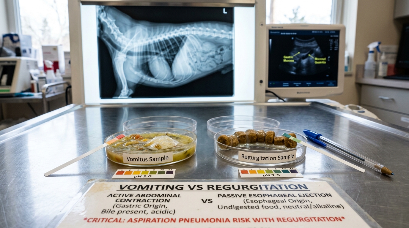
The core difference between dog vomiting and regurgitation is active abdominal effort. Vomiting is an active reflex preceded by nausea, lip-licking, and forceful abdominal heaving that expels digested food and yellow-green bile from the stomach. Regurgitation is passive, effortless expulsion where undigested food drops out of the esophagus with zero abdominal contraction.
Clinical Quick Answer & Immediate Actions
Distinguishing vomiting from regurgitation is a vital clinical crossroad. Vomiting is an active, reflexive gastrointestinal process preceded by nausea (lip-licking, hypersalivation) and characterized by forceful abdominal heaving that expels digested or partially digested food, bile, or gastric fluids. In contrast, regurgitation is a passive, effortless esophageal failure where undigested food, saliva, or mucus is ejected retrograde without preceding abdominal contraction or warning. Regurgitation indicates esophageal dysfunction (such as megaesophagus, vascular ring anomaly, or foreign body obstruction) and carries an acute risk of life-threatening aspiration pneumonia.
What Is the Difference Between Dog Vomiting and Regurgitation?
Vomiting is an active gastric reflex with forceful retching and bile expulsion. Regurgitation is passive esophageal release with no retching or bile. While occasional mild vomiting may respond to temporary fasting, persistent regurgitation is a veterinary emergency carrying high risk of aspiration pneumonia.
When a dog brings up food or liquid, pet owners routinely tell the triage veterinary team that their pet has “thrown up.” However, veterinary internal medicine makes a fundamental distinction between active vomiting and passive regurgitation. Confusing these two clinical presentations can lead to misdirected diagnostics, delayed emergency surgery, or life-threatening complications such as aspiration pneumonia. this detailed clinical guide outlines the physiological mechanisms, diagnostic comparison metrics, MSD triage emergency red flags, and evidence-based home observation protocols.
Neurophysiology: Active Vomiting vs. Passive Regurgitation
Understanding the anatomical pathways of these two phenomena explains why their clinical management is radically different:
1. Active Vomiting (Gastric & Upper Duodenal Origin)
Vomiting is a multi-step neurochemical reflex orchestrated by the vomiting center in the medulla oblongata, triggered either via bloodborne toxins stimulating the chemoreceptor trigger zone (CRTZ) in the area postrema, or via visceral afferent sympathetic and vagal pathways from the gastrointestinal tract, peritoneum, or vestibular apparatus. The process follows three distinct clinical phases:
- Prodromal (Nausea) Phase: Mediated by autonomic stimulation, manifesting as intense restlessness, excessive swallowing, ptyalism (drooling), lip smacking, and tachypnea.
- Retching Phase: Involuntary, coordinated spasmodic contractions of the diaphragm, abdominal wall musculature, and thoracic wall occur while the gastroesophageal junction and glottis remain tightly closed, generating negative intrathoracic and positive intra-abdominal pressure.
- Expulsion Phase: The gastroesophageal sphincter relaxes, the stomach contracts, the soft palate elevates to close the nasopharynx, and the gastric contents are forcibly ejected out of the oral cavity.
2. Passive Regurgitation (Esophageal Origin)
Unlike vomiting, regurgitation involves no central nervous reflex coordination, no prodromal nausea, and no abdominal musculature involvement. Food, fluid, or saliva that fails to reach the stomach accumulates within the lumen of the esophagus due to mechanical obstruction, motility failure, or lower esophageal sphincter dysfunction. When the animal lowers its head or gravity overcomes esophageal resistance, the accumulated contents passively roll forward and discharge from the mouth. The expelled material is unacidified (neutral to alkaline pH) and has never been exposed to gastric enzymes or duodenal bile.
Diagnostic Comparison Matrix
The following clinical triage matrix highlights the key differentiating parameters evaluated by veterinary staff during initial patient intake:
| Diagnostic Indicator | Active Vomiting (Stomach/Intestines) | Passive Regurgitation (Esophagus) |
|---|---|---|
| Prodromal Warning Signs | Present: restless pacing, heavy drooling, lip smacking, frequent swallowing. | Absent: pet acts normal right until the moment food spills out. |
| Abdominal Effort & Sound | Violent abdominal heaving, rhythmic dry retching, arched posture. | Zero abdominal contraction; completely silent and effortless. |
| Expelled Material Appearance | Partially digested, acidic smell, frequent yellow/green bile or gastric foam. | Undigested, tubular cylinder shape, covered in thick clear/white mucus. Zero bile. |
| Chemical pH Level | Acidic (pH < 5.0) due to hydrochloric gastric acid secretion. | Neutral to alkaline (pH 7.0–8.0) from salivary and esophageal secretions. |
| Primary Etiologies | Dietary indiscretion, GDV, pancreatitis, foreign body, renal failure, toxins. | Megaesophagus, Myasthenia Gravis, esophageal foreign body, stricture, PRAA. |
| Secondary Critical Risk | Severe hypokalemia, hypochloremic alkalosis, hypovolemic shock. | Fatal aspiration pneumonia from inhalation of esophageal fluids. |
Emergency Red Flags: When to Seek Urgent Veterinary Care
Based on emergency triage protocols outlined by the MSD Veterinary Manual and the American College of Veterinary Emergency and Critical Care (VECCS), the following signs indicate a medical emergency where patient survival depends on prompt intervention:
1. Gastric Dilatation-Volvulus (GDV / Bloat) — Surgical Emergency
GDV is a hyperacute condition where the stomach distends with gas and rotates along its mesenteric axis. It rapidly occludes venous return via the caudal vena cava, inducing severe cardiogenic and obstructive shock:
- Hallmark Signs: Continuous, non-productive retching (trying to vomit but nothing comes out), rapid abdominal distension that sounds tympanic (drum-like) upon gentle percussion, severe restlessness, and praying posture.
- Clinical Emergency Stabilization: Immediate dual large-bore IV catheter placement (18-gauge in cephalic or jugular veins) for shock-dose fluid resuscitation (10–prescribed clinical dosage isotonic crystalloids over 15 minutes), prompt gastric decompression via orogastric tube or percutaneous trocarization with an 18-gauge needle at the point of maximal tympany, ECG monitoring for ventricular premature contractions (VPCs), and emergency surgical gastropexy within 1–2 hours.
2. Severe Gastrointestinal Hemorrhage (Hematemesis & Melena)
The presence of fresh frank blood or dark brown granular material resembling “coffee grounds” in the vomit (hematemesis) indicates acute erosive gastroduodenitis or ulcerative perforation. Ingested blood that digests further along the intestine produces black, sticky, tarry stools (melena). Frequent causes include indiscriminate use of human non-steroidal anti-inflammatory drugs (NSAIDs) or concurrent corticosteroid administration. Perforating ulcers risk pneumoperitoneum and fatal septic peritonitis.
3. Circulatory Shock & Vital Sign Deterioration
Evaluate your dog’s vital perfusion signs using the home triage method:
- Mucous Membrane Color: Lift the upper lip to inspect the gums. Pale, porcelain-white, grayish, or cyanotic (bluish) gums signify hypovolemic shock, internal hemorrhage, or respiratory collapse. Injected brick-red gums indicate severe systemic inflammatory response (SIRS) or sepsis.
- Capillary Refill Time (CRT): Press firmly on the pink gum tissue and release. Perfusion should return within 1.5 to 2.0 seconds. A CRT exceeding 2.0 seconds indicates peripheral vasoconstriction and severe circulatory compromise; a CRT under 1.0 second indicates hyperdynamic shock.
- Pulse Quality & Heart Rate: Tachycardia (> 140–160 bpm in medium-to-large dogs) or life-threatening bradycardia (< 60 bpm) accompanied by weak femoral pulses signals decompensated shock or dangerous hyperkalemia.
4. Electrolyte and Acid-Base Derangements
Clinical studies (such as research published in SciELO on canine gastric conditions) demonstrate that protracted vomiting results in hypokalemia in 72.1% of clinical canine presentations, alongside hypochloremia and metabolic alkalosis caused by gastric hydrochloric acid depletion. Severe potassium depletion impairs myocardial conductivity and induces neuromuscular weakness, leaving the pet collapsed and unable to stand.
5. Secondary Aspiration Pneumonia
Dogs suffering from persistent regurgitation (especially those diagnosed with acquired Myasthenia Gravis or congenital megaesophagus) are at acute risk of inhaling esophageal contents into the bronchial tree. Warning signs include moist coughing, raspy crackles on thoracic auscultation, persistent purulent nasal discharge, lethargy, and a body temperature exceeding 39.5°C (103.1°F). This complication carries a high mortality rate without immediate oxygen supplementation, systemic broad-spectrum IV antibiotics, and nebulization therapy.
Evidence-Based At-Home Care Protocol
If your adult dog has experienced a single, isolated mild vomiting episode, remains fully alert, displays pink gums, and shows zero signs of pain or abdominal enlargement, you may implement this conservative home care pathway:
- Temporary Food Withholding (6–8 Hours): Allow the irritated gastric mucosal lining to rest. Do not offer kibble, canned food, or rich treats during this period.
- Controlled Micro-Hydration: Gulping large volumes of cold water triggers gastric stretch receptors, causing immediate rebound vomiting. Instead, offer 1 to 2 tablespoons of room-temperature water or ice cubes every 30 to 45 minutes to maintain hydration without overfilling the stomach.
- Gradual Bland Diet Introduction: If no further vomiting occurs after 8 hours, offer a small meal (1–2 tablespoons) composed of boiled skinless chicken breast or lean ground beef mixed with boiled white rice in a 1:2 ratio. Feed small, frequent meals 3–4 times daily for 48 hours before gradually transitioning back to standard commercial food over 3–5 days.
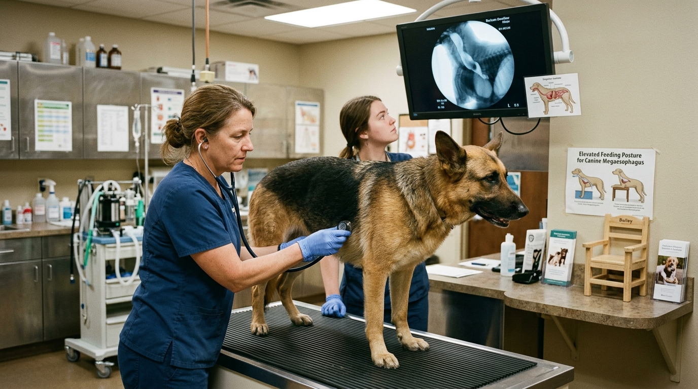
- Postural Management for Regurgitation: If regurgitation is suspected, never feed the dog flat on the floor. Use an elevated feeding bowl or a specialized Bailey Chair that maintains the pet in an upright vertical position for 20 to 30 minutes post-feeding, allowing gravity to facilitate passage of food into the stomach.
- Absolute Contraindication: Never administer human over-the-counter anti-nausea drugs, aspirin, paracetamol (acetaminophen), or ibuprofen. These compounds are severely toxic to canines and trigger massive gastric perforation, acute renal shutdown, and liver necrosis. Contact your licensed veterinary clinic immediately if symptoms persist beyond 24 hours.
Clinical Practice Tool: Veterinary Preparation
When preparing for a clinical evaluation of acute or chronic vomiting, download our free printable Veterinary Visit Preparation Sheet to log precise timelines, fluid retention, and vital signs for your clinician. Browse all clinical templates in the NewsPet Resource Center.
Frequently Asked Questions
Why does my dog vomit yellow foam early in the morning?
Early morning yellow foam vomiting is classic Bilious Vomiting Syndrome (BVS). When the stomach remains empty for an extended period, duodenal motility allows intestinal bile to reflux backward across the pyloric sphincter into the stomach, irritating the mucosa. Feeding a small protein-rich snack or high-fiber biscuit right before bedtime often eliminates this condition.
Can eating grass cause my dog to vomit or regurgitate?
Ingesting fibrous grass blades mechanically irritates the gastric lining and tickles the pharyngeal mucosa, triggering active vomiting rather than regurgitation. While occasional grass eating is common canine behavior, compulsive ingestion coupled with retching warrants an examination for underlying inflammatory bowel disease or chronic gastric hyperacidity.
What diagnostic tests will the vet perform?
Your veterinarian will likely perform physical abdominal palpation, complete blood count (CBC) and serum biochemistry (evaluating BUN, creatinine, ALT, lipase, and electrolyte levels), and plain or contrast thoracic and abdominal radiographs. X-rays are crucial to differentiate between an esophageal megaesophagus dilation and gastric foreign bodies or obstructive ileus.
Frequently Asked Questions
How can I tell if my dog is vomiting or regurgitating?
Vomiting is an active process preceded by nausea, lip-licking, pacing, and forceful abdominal heaving that expels partially digested food, yellow bile, or foam. Regurgitation is a passive, effortless event with zero abdominal heaving where undigested, tubular food coated in clear saliva drops out of the mouth shortly after eating.
Is regurgitation in dogs a veterinary emergency?
Yes, persistent or sudden regurgitation requires urgent veterinary examination. While vomiting originates in the stomach, regurgitation points to esophageal disorders like megaesophagus, foreign body obstruction, or vascular ring anomalies, and carries a high risk of life-threatening aspiration pneumonia.
What should I do at home if my dog regurgitates food?
Withhold further food and water for 2 hours to prevent acute aspiration into the lungs, document the appearance and timing of the expelled food, check your dog’s breathing rate, and contact your veterinarian. Never administer human stomach medications or force liquids into a regurgitating dog.
Sources
- CABI Digital Library: Differentiating Vomiting from Regurgitation in Dogs (opens in new tab)
- MSD Veterinary Manual: Triage Parameters & Gastrointestinal Pathology in Small Animals (opens in new tab)
- VCA Animal Hospitals: Clinical Evaluation and Management of Canine Megaesophagus and Myasthenia Gravis (opens in new tab)
- SciELO Veterinary Research: Electrolyte and Acid-Base Imbalances in Dogs with Hemorrhagic Gastroenteritis (opens in new tab)
- DVM360: Clinical Emergency Stabilization Protocol for Gastric Dilatation-Volvulus (GDV) (opens in new tab)
Emergency Triage Matrix: Clinical Presentation & Required Response
| Triage Level | Clinical Signs & Presentation | Immediate Action Protocol |
|---|---|---|
| Tier 1: Immediate Emergency (0–2 Hours) | Regurgitation or vomiting accompanied by coughing, crackling lung sounds, fever >103.5°F, dyspnea, or cyanosis (aspiration pneumonia); or active vomiting with unproductive retching, distended rock-hard abdomen (GDV/bloat), or coffee-ground hematemesis. | Immediate emergency veterinary hospitalization; oxygen therapy, thoracic radiography, intravenous fluid stabilization, suction airway clearance, and injectable broad-spectrum antimicrobial or surgical intervention. |
| Tier 2: Urgent Veterinary Care (Same-Day) | Frequent daily regurgitation of undigested tubular food boluses within minutes of eating; persistent regurgitation with progressive weight loss; or repeated vomiting >3 episodes in 24 hours with mild dehydration. | Same-day veterinary evaluation; contrast fluoroscopy, thoracic radiographs to evaluate esophageal lumen dilation, complete blood chemistry, and baseline gastrointestinal workup. |
| Tier 3: Home Monitoring & Scheduled Care | Single episode of regurgitation after rapid eating/gulping dry kibble in an otherwise energetic dog with clear lungs, normal respiration, and pink mucous membranes. | Transition to slow-feeder bowls, elevate food bowl slightly, feed smaller divided meals, and monitor resting respiratory rate while sleeping for any signs of early aspiration. |
Rule S1 Boundaries & Esophageal Safety Rules
Esophageal and gastrointestinal disorders require strict physical and pharmacological boundaries:
- NEVER force water or broth immediately following regurgitation: The laryngeal protective reflex is compromised; oral fluids can flow directly into the trachea, triggering catastrophic acute aspiration pneumonia and asphyxiation.
- NEVER induce vomiting in a dog suspected of esophageal obstruction: Forcing caustic contents or sharp foreign objects back up through an inflamed or compromised esophagus risks full-thickness esophageal perforation and mediastinitis.
- NEVER feed a megaesophagus patient in a flat, horizontal posture: Canines with dilated megaesophagus lack peristaltic motility; meals must be administered in a vertical 90-degree position (e.g., Bailey chair) maintained for 20–30 minutes post-prandially to utilize gravity.
- NEVER administer human antiemetics or acid reducers without radiographic clearance: Suppressing nausea with over-the-counter pharmaceuticals masks mechanical foreign bodies or complete intestinal obstructions, leading to bowel necrosis.
Caregiver Clinical Action Checklist: Vomiting vs. Regurgitation
- Capture high-definition video of the expulsion episode showing the pet’s abdomen and posture to present to your veterinarian.
- Inspect the expelled material: note whether food is undigested and tubular (esophageal) or digested with yellow/green bile (gastric).
- Count resting respiratory rate (RRR) while the pet is sleeping; verify breaths remain below 30 breaths per minute.
- Assess gum color and capillary refill time; ensure membranes remain healthy salmon pink and refill within 1.5 seconds.
- Place food and water bowls on an elevated platform pending formal veterinary thoracic radiography.
- Transport immediately to an emergency facility if coughing, wet lung rattles, or fever develop.
Related Clinical Guides & Tools
Peer-Reviewed Clinical References & Evidence Grounding
- PMID: 33769611: International veterinary canine dyskinesia task force ECVN consensus statement: Terminology and classification (J Vet Intern Med, 2021) (opens in new tab)
- PMID: 38351524: Defining sepsis in small animals (J Vet Emerg Crit Care (San Antonio), 2024) (opens in new tab)
- PMID: 38474142: Canine Mammary Tumors: Classification, Biomarkers, Traditional and Personalized Therapies (Int J Mol Sci, 2024) (opens in new tab)
- Merck Veterinary Manual — Small Animal Clinical Medicine & Emergency Reference (opens in new tab)
Pet Toxic Food & Hazard Fast-Checker
Rapidly verify toxicity risks, toxic mechanisms, and immediate clinical steps for common household foods, plants, and human medications before calling poison control or your veterinarian.
Printable tools
Download the matching worksheet
Source transparency
Reference mix for this guide
Source labels describe the type of organization behind each reference; they are not a claim that any outside organization reviewed this article.
- CABI Digital Library: Differentiating Vomiting from Regurgitation in Dogs Reference source · cabidigitallibrary.org
- MSD Veterinary Manual: Triage Parameters & Gastrointestinal Pathology in Small Animals Veterinary organization · merckvetmanual.com
- VCA Animal Hospitals: Clinical Evaluation and Management of Canine Megaesophagus and Myasthenia Gravis Reference source · vcahospitals.com
- SciELO Veterinary Research: Electrolyte and Acid-Base Imbalances in Dogs with Hemorrhagic Gastroenteritis Veterinary organization · scielo.br
- DVM360: Clinical Emergency Stabilization Protocol for Gastric Dilatation-Volvulus (GDV) Reference source · dvm360.com
- Puppy Socialization Without Overwhelm: A Vet-Safe Reference source · newspet.net
- Dog Body Language Chart: 9 Stress Signs Before a B Reference source · newspet.net
- Reward-Based Dog Training: Force-Free Steps & Reference source · newspet.net