This guide is general education for pet owners. If your pet has urgent symptoms, possible poisoning, injury, breathing trouble, collapse, severe pain, or sudden decline, contact a licensed veterinarian or local emergency service.
Dosages must be calculated by a licensed veterinarian based on species, weight, and bloodwork. Never administer human medications or self-adjust prescription doses for pets.
Editorial standard
How this guide was prepared
Septic peritonitis represents the pinnacle of surgical emergencies in veterinary critical care, defined by acute, widespread microbial contamination and life-threatening inflammation of the peritoneal cavity. In dogs, this catastrophic condition almost invariably results from the physical rupture or perforation of an internal abdominal organ—most frequently the gastrointestinal tract due to foreign body ulceration, severe necrotizing pancreatitis, or dehiscence of a previous surgical incision. Less commonly, it arises from the rupture of an infected pyometra, an infected prostatic abscess, or a compromised gallbladder with infected bile peritonitis. When millions of enteric bacteria and endotoxins spill into the sterile peritoneal space, the vast surface area of the peritoneum absorbs these toxins into the general circulation, precipitating massive Systemic Inflammatory Response Syndrome (SIRS), profound vasodilation, refractory septic shock, and multi-organ dysfunction syndrome (MODS). Survival depends on immediate recognition, rapid point-of-care diagnostics, aggressive cardiovascular stabilization, and emergency surgical source control.
Quick Answer: Recognizing Acute Abdomen & Septic Peritonitis in Dogs
If your dog suddenly exhibits a rigid, painful “board-like” abdomen, adopts the “prayer position” (front paws down, rear end elevated in pain), develops pale or muddy brick-red gums, vomits relentlessly, or collapses with rapid, shallow breathing, suspect acute septic peritonitis from a ruptured internal organ. This condition cannot be treated at home. Transport your dog immediately to an emergency hospital. Emergency clinicians perform a focused ultrasound (AFAST) and bedside abdominocentesis (fluid tap). If peritoneal fluid shows intracellular bacteria under the microscope, or if the fluid glucose is >20 mg/dL lower than blood glucose, immediate emergency surgical laparotomy, extensive peritoneal lavage, and intensive care hospitalization are required.
Etiology: What Causes Organs to Rupture in the Canine Abdomen?
The peritoneal cavity is a delicate, sterile space lined by a single layer of mesothelial cells with an immense absorptive surface. Septic peritonitis occurs when this barrier is violated, leading to overwhelming bacterial colonization:
1. Gastrointestinal Perforation (50–70% of cases): Sharp or linear foreign bodies (bones, sticks, needles), deep perforating peptic or neoplastic ulcers, or mechanical bowel strangulation (such as untreated volvulus or intussusception) cause full-thickness bowel necrosis. Another frequent cause is surgical anastomotic dehiscence 3 to 5 days following prior intestinal surgery.
2. Hepatobiliary Rupture (Bile Peritonitis): Ruptured gallbladder mucoceles, severe necrotizing cholecystitis, or biliary trauma allow caustic bile salts to flood the peritoneum. When bile is contaminated with ascending bacteria, chemical peritonitis rapidly converts to fulminant septic shock.
3. Urogenital Rupture: Ruptured closed-cervix pyometra, ruptured infected prostatic abscesses, or infected urinary bladder trauma allow purulent fluid or infected urine (uroabdomen with secondary sepsis) to trigger massive bacteremia.
4. Penetrating Abdominal Trauma: Bite wounds from larger dogs, vehicular trauma, or penetrating impalement injuries drive external environmental bacteria deep into the peritoneal cavity.
Clinical Emergency Triage Matrix
| Triage Tier | Clinical Presentation | Required Action |
|---|---|---|
| Tier 1: Septic Shock & Acute Peritoneal Crisis (0-1h) | Profound collapse or stupor; rigid, tense “board-like” abdomen that cries out or growls upon gentle touch; “prayer position” posture; body temperature <37.2°C (<99°F, decompensated septic shock) or >40°C (>104°F); brick-red “injected” or grayish-white gums; capillary refill time >3 seconds or instantaneous (<1 sec); weak thready femoral pulses. | Proceed immediately to the nearest 24-hour veterinary ICU hospital. Withhold all oral intake. Telephone the emergency hospital while traveling so the trauma team can prepare oxygen support, multiple large-bore IV catheters, balanced resuscitation fluids, broad-spectrum IV antimicrobials, and an operating suite for emergency laparotomy. |
| Tier 2: Progressive Distension & Persistent Emesis (1-4h) | Rapidly enlarging, distended abdomen with a distinct “fluid wave”; persistent vomiting of bile or foul-smelling fluid; intense restlessness, inability to get comfortable lying down; history of recent abdominal surgery 3-5 days ago. | Transport immediately to an emergency veterinary clinic. Rapid bedside ultrasound (AFAST) and abdominal fluid analysis must be performed without delay to detect early anastomotic leakage or localized abscess rupture before cardiovascular collapse ensues. |
| Tier 3: Moderate Abdominal Guarding & Lethargy (4-12h) | Mild abdominal guarding and discomfort when walking; reluctance to jump; subdued appetite and mild dehydration; normal gum color and stable pulse quality. | Schedule an urgent same-day veterinary consultation. Perform a thorough physical examination, abdominal survey radiographs, and complete blood count with chemistry panel to investigate early inflammatory abdominal pathology. |
Dangerous Home Pitfalls: What NOT to Do (Rule S1)
- NEVER administer human pain relievers (ibuprofen, naproxen, paracetamol/acetaminophen): Human NSAIDs and acetaminophen are profoundly toxic to dogs. In a dog suffering from peritonitis, they cause devastating gastric perforations, accelerate acute kidney shutdown, and cause fatal liver necrosis.
- NEVER attempt home needle drainage (paracentesis) of an enlarged abdomen: Puncturing the abdominal wall with a needle or syringe without ultrasound guidance risks lacerating friable intestines, the spleen, or the urinary bladder, introducing further contamination.
- NEVER offer food, milk, or oral electrolytes: Ingested material entering a perforated stomach or bowel will directly escape into the peritoneal cavity, doubling the bacterial load and guaranteeing lethal peritonitis.
- NEVER apply heating pads directly to a pet in septic shock: Direct external heat induces peripheral cutaneous vasodilation, shunting blood away from vital organs (brain, heart, kidneys) and worsening fatal hypovolemic hypotension.
Definitive Bedside Diagnostics: The Glucose-Lactate Gradient
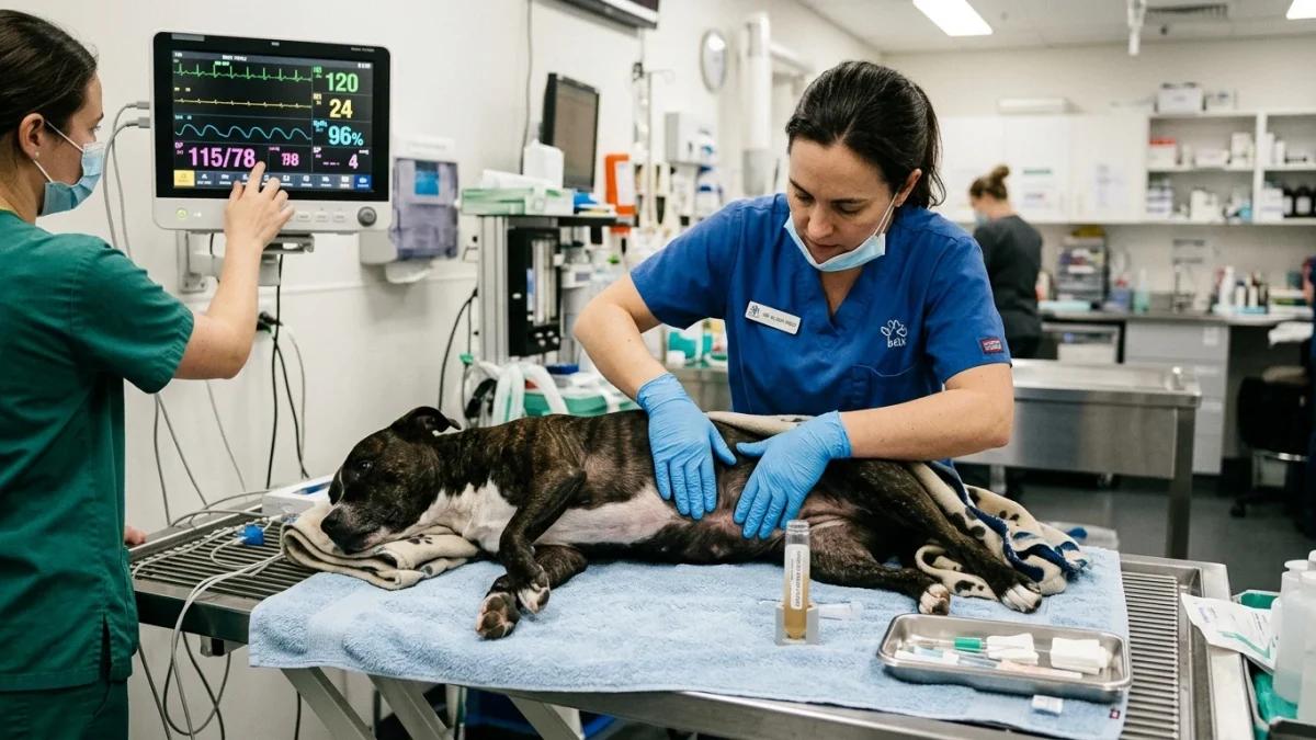
When an emergency patient arrives with an acute abdomen, the veterinary team conducts an emergency Focused Assessment with Sonography for Trauma (AFAST) to scan four acoustic windows for peritoneal free fluid (diaphragmatic-hepatic, splenorenal, cystocolic, and hepatorenal).
If free fluid is detected, an immediate bedside abdominocentesis is performed. Definitive diagnosis of septic peritonitis relies on two rapid point-of-care modalities:
1. Cytological Examination: A direct smear of the fluid is stained and examined under oil immersion. The hallmark finding is the presence of intracellular bacteria (cocci or rods) engulfed within the cytoplasm of degenerate, toxic neutrophils.
2. The Glucose and Lactate Gradient (Diagnostic Standard): Rapid biochemical testing using bedside point-of-care glucometers and lactate meters compares abdominal fluid to peripheral blood:
- Fluid Glucose Difference: A peritoneal fluid glucose concentration that is more than 20 mg/dL lower than blood glucose indicates active, heavy bacterial metabolism and leukocyte phagocytosis, diagnosing septic peritonitis with 100% sensitivity.
- Fluid Lactate Difference: A peritoneal fluid lactate concentration that is more than 2.0 mmol/L higher than peripheral blood lactate demonstrates anaerobic peritoneal tissue hypoperfusion and severe local sepsis.
Emergency Stabilization and Surgical Source Control
Septic peritonitis cannot be cured with medications alone; it requires surgical elimination of the source of contamination. However, rushing an unstable, hypotensive patient directly into surgery carries an unacceptably high anesthetic mortality. Emergency clinicians follow a structured resuscitation protocol:
Phase 1: Hemodynamic Resuscitation: Rapid intravenous crystalloid fluid resuscitation restores effective circulating volume, accompanied by continuous ECG and blood pressure monitoring. Broad-spectrum four-quadrant intravenous antibiotics (such as ampicillin-sulbactam combined with enrofloxacin or a fluoroquinolone) are administered immediately after fluid collection.
Phase 2: Surgical Debridement & Repair: Through a full-length ventral midline exploratory celiotomy, the surgeon systematically locates the perforation, resects devitalized intestinal segments, performs closure with leak testing, and removes all purulent debris.
Phase 3: Copious Peritoneal Lavage: The entire abdominal cavity is lavaged with several liters of warm, sterile isotonic saline (often extensive multi-liter volumes proportional to the dog’s body size) until the returned fluid is crystal clear, followed by suction evacuation. Depending on the severity of contamination, an active closed-suction drain (Jackson-Pratt) is placed.
Frequently Asked Questions
What is the typical survival rate for dogs with septic peritonitis?
Published survival rates for dogs treated with emergency surgical debridement and intensive post-operative ICU care range between 50% and 70%. Factors that significantly reduce survival include delayed presentation, refractory septic shock, multiple organ failure, and underlying neoplastic rupture. Early diagnosis within the first few hours of perforation dramatically increases the likelihood of a full recovery.
Why does an infected abdomen make a dog’s gums look brick red?
In the hyperdynamic early phase of septic shock, bacterial endotoxins trigger widespread, uncontrolled release of nitric oxide and inflammatory cytokines, causing massive peripheral vasodilation. Capillaries in the mucous membranes become engorged with blood, creating an intense “brick-red” or “injected” appearance with a bounding pulse and instantaneous capillary refill time (<1 second), which subsequently collapses into grayish-pale gums as shock progresses.
How can I tell if my dog’s prior surgery is breaking down internally?
Dehiscence of gastrointestinal incisions most commonly occurs between Day 3 and Day 5 post-operatively, coinciding with the lag phase of wound healing where fibrin seals dissolve before new collagen gains tensile strength. Warning signs include a sudden relapse into lethargy, refusal to eat, low-grade fever, vomiting, abdominal guarding, and fluid swelling along the ventral incision line.
Scientific Sources and Literature
- Bonczynski JJ, et al. “Comparison of peritoneal fluid and peripheral blood glucose and lactate concentrations in dogs and cats with septic peritonitis.” Journal of the American Veterinary Medical Association. PMID: 29774621
- Culp WT, et al. “Septic peritonitis in dogs: pathophysiology, diagnosis, and stabilization prior to surgery.” Veterinary Clinics of North America: Small Animal Practice. PMID: 26031454
- Mueller MG, et al. “Prognostic factors, surgical techniques, and open versus closed peritoneal drainage in canine septic peritonitis: 110 cases.” Journal of Veterinary Emergency and Critical Care. PMID: 32304192
Caregiver Emergency Acute Abdomen Action Checklist
- Assess abdominal posture: note if the dog guards its belly, arches its back in the “prayer position”, or cries when touched.
- Check gum color: observe whether gums are brick red (early sepsis) or white/muddy gray (decompensated shock).
- Do not administer any human medications, NSAIDs, or aspirin under any circumstances.
- Never attempt to insert needles or extract abdominal fluid at home.
- Do not offer food, milk, water, or oral electrolytes; keep the stomach completely empty for emergency surgery.
- Keep the dog wrapped in a warm dry blanket during car transport, avoiding direct electrical heating pads.
- Proceed directly to a 24-hour veterinary emergency center equipped with ultrasound and surgical intensive care.
Source transparency
Reference mix for this guide
Source labels describe the type of organization behind each reference; they are not a claim that any outside organization reviewed this article.
- Dog Sudden Collapse & Internal Bleeding Triage Reference source · newspet.net
- Dog Acute Pancreatitis & Prayer Position Protocol Reference source · newspet.net
- Dog Pyometra Uterine Infection Triage Reference source · newspet.net
- Dog Surgical Incision Healing vs Infection Triage Reference source · newspet.net
- PMID: 29774621 Government or public agency · pubmed.ncbi.nlm.nih.gov
- PMID: 26031454 Government or public agency · pubmed.ncbi.nlm.nih.gov
- PMID: 32304192 Government or public agency · pubmed.ncbi.nlm.nih.gov