This guide is general education for pet owners. If your pet has urgent symptoms, possible poisoning, injury, breathing trouble, collapse, severe pain, or sudden decline, contact a licensed veterinarian or local emergency service.
Dosages must be calculated by a licensed veterinarian based on species, weight, and bloodwork. Never administer human medications or self-adjust prescription doses for pets.
Editorial standard
How this guide was prepared
Canine Infectious Respiratory Disease Complex (CIRDC), historically termed kennel cough, is not a single-pathogen illness but a highly contagious, multifactorial respiratory syndrome driven by synergistic viral and bacterial co-infections. Primary viral agents—including canine parainfluenza virus (CPIV), canine adenovirus-2 (CAV-2), canine respiratory coronavirus (CRCoV), and canine influenza virus (CIV H3N8 and H3N2)—denude the respiratory epithelium, enabling bacterial colonizers such as Bordetella bronchiseptica, Mycoplasma cynos, and Streptococcus equi subsp. zooepidemicus to establish deep tracheobronchial or alveolar pathology.
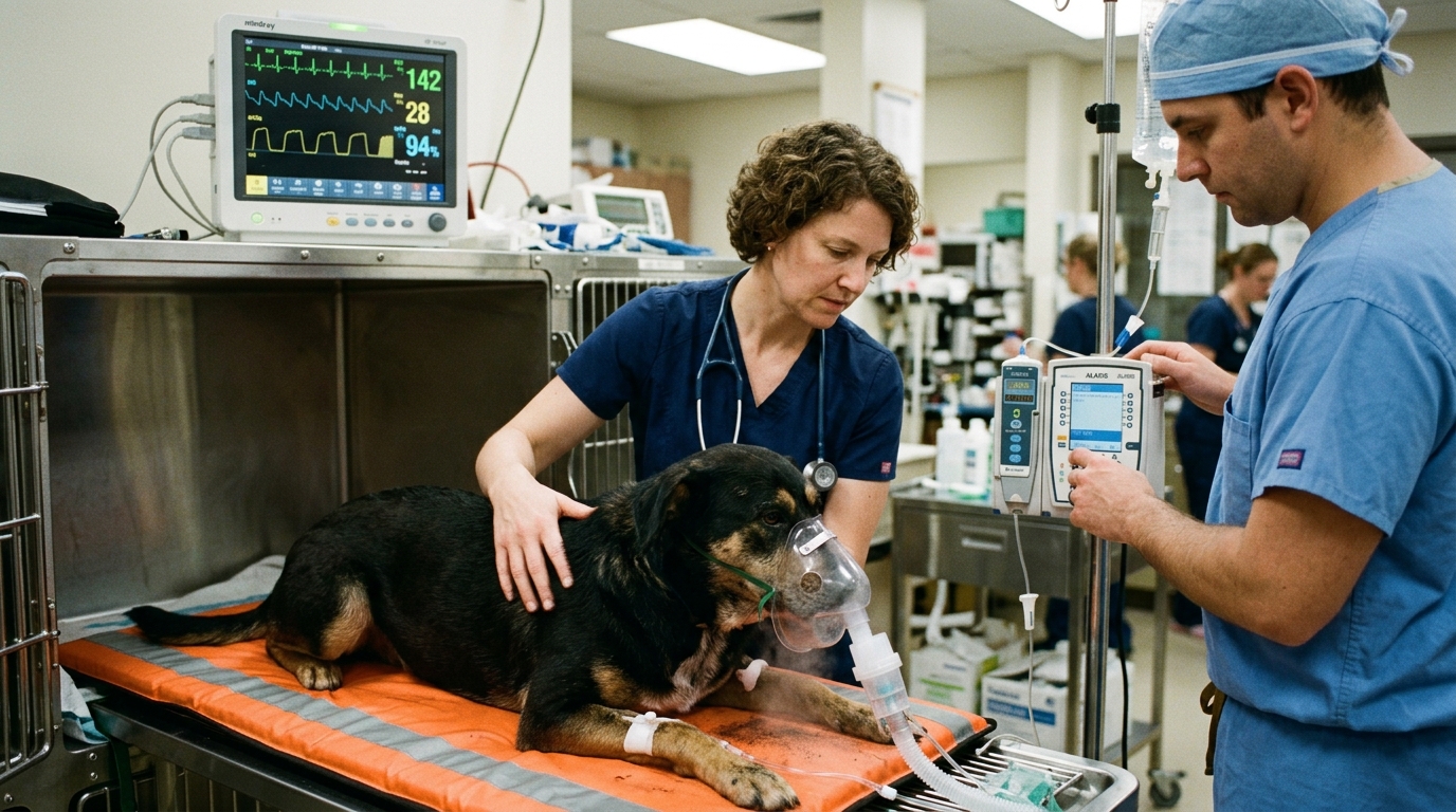
Clinical Quick Answer & Immediate Stabilization Protocol
Primary Clinical Directive for Canine Infectious Respiratory Disease Complex (CIRDC) & Secondary Bacterial Bronchopneumonia: A sudden, paroxysmal “goose-honk” dry cough ending in a terminal white frothy gag in an otherwise bright, afebrile dog typically reflects upper airway tracheobronchitis following daycare, boarding, or grooming exposure. However, if the cough becomes soft and moist accompanied by fever (>103.0°F), lethargy, loss of appetite, yellow-green nasal exudate, or increased abdominal breathing effort, the infection has likely descended into the lung parenchyma. Follow ISCAID antimicrobial stewardship guidelines by avoiding empirical antibiotics in mild self-limiting cases while seeking rapid thoracic radiography and targeted therapy when pneumonia indicators emerge.
Ciliostasis, Epithelial Desquamation & Polymicrobial Synergy
The healthy canine tracheobronchial tree relies on the mucociliary escalator—a coordinated blanket of ciliated pseudostratified columnar epithelium and mucus-secreting goblet cells—to trap and expel inhaled particulates before they reach the sterile alveoli. Viral pathogens like canine parainfluenza and canine influenza replicate within these respiratory epithelial cells, causing widespread desquamation and exposing basement membrane collagen within 48 to 72 hours.
Concurrently, Bordetella bronchiseptica expresses filamentous hemagglutinin, pertactin, and fimbrial adhesins that bind directly to respiratory cilia, secreting tracheal cytotoxin and adenylate cyclase hemolysin. These bacterial exotoxins paralyze ciliary beat frequency (ciliostasis) within 3 hours of attachment and inhibit neutrophil phagocytosis. When mucus stagnates in the lower airways, opportunistic organisms such as Mycoplasma cynos and enteric gram-negative rods proliferate in the cranioventral lung lobes, transforming a dry tracheal irritation into suppurative bronchopneumonia.
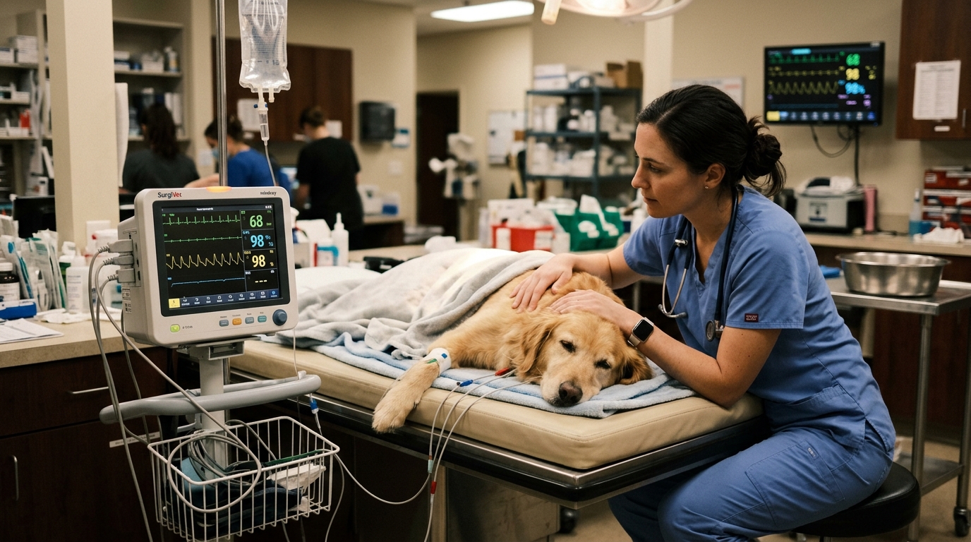
- Tracheal Hypersensitivity & Terminal Gag: Exposed subepithelial vagal nerve endings in the larynx and trachea trigger violent paroxysms of coughing upon minimal collar pressure or cold air inhalation, frequently culminating in expectoration of white tracheobronchial foam mistaken by owners for vomit.
- Canine Influenza H3N2 Shedding Window: Unlike CPIV or CAV-2 (which shed for 5–7 days), H3N2 canine influenza can be shed intermittently in nasal secretions for up to 21 to 26 days, requiring extended 4-week household isolation.
- Streptococcus zooepidemicus Hemorrhagic Pneumonia: Co-infection with S. equi subsp. zooepidemicus triggers superantigen-mediated cytokine storms, causing peracute necrohemorrhagic pneumonia and pleural effusion.
- Brachycephalic & Tracheal Collapse Predisposition: Pugs, French Bulldogs, and toy breeds with dynamic airway collapse experience severe turbulent airflow and impaired clearance, elevating their risk of rapid respiratory decompensation.
Emergency Triage Matrix: Clinical Presentation & Required Response
| Triage Level | Clinical Signs & Presentation | Immediate Action Protocol |
|---|---|---|
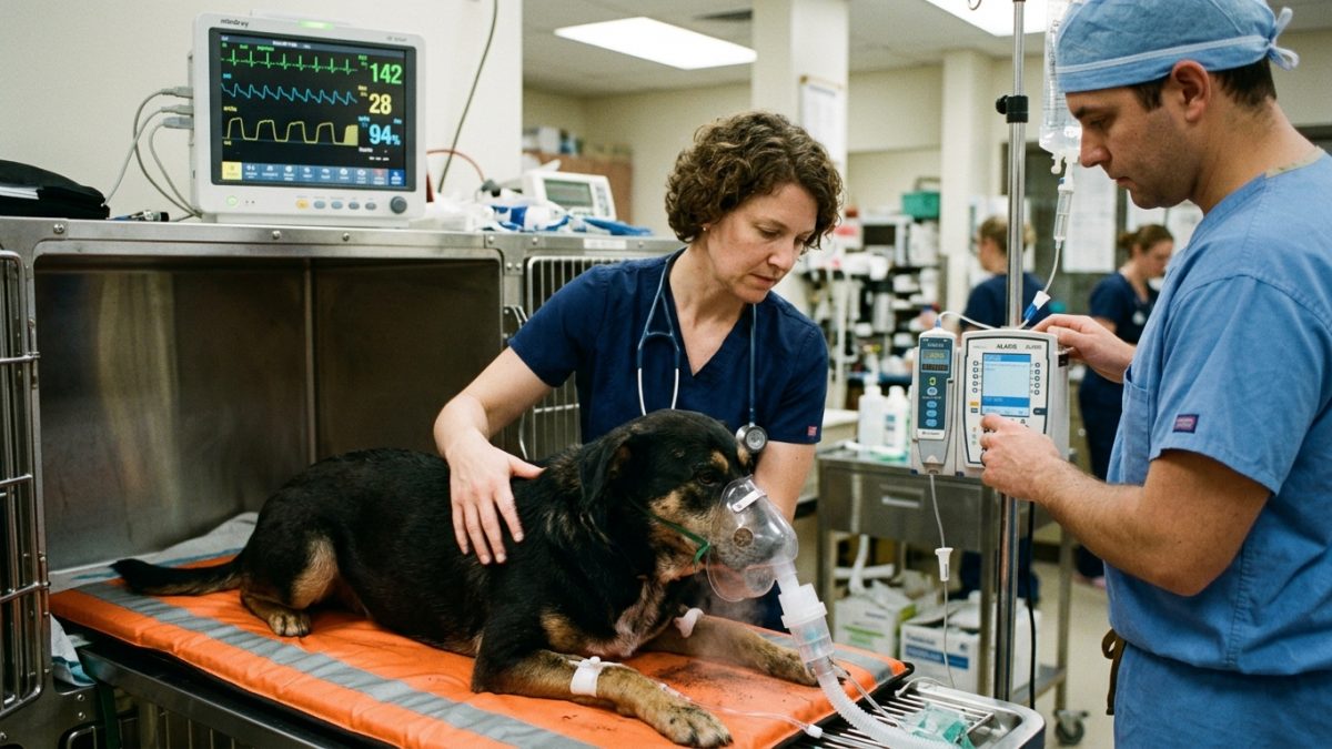
| Tier 1: Immediate Emergency (0–1 Hour) | Resting respiratory rate >40 breaths/min, extended neck with elbows abducted (orthopnea), cyanotic or pale gums, hemoptysis (coughing blood), rectal temperature >103.5°F, or profound collapse. | Transport immediately to an emergency veterinary facility for flow-by or cage oxygen supplementation, three-view thoracic radiographs, pulse oximetry, and parenteral bactericidal antimicrobials. |
| Tier 2: Urgent Same-Day Evaluation (2–12 Hours) | Transition from dry honking cough to moist productive cough, thick mucopurulent yellow/green nasal or ocular discharge, refusal of meals, lethargy, or persistent cough in a puppy, senior, or brachycephalic dog. | Arrange a same-day veterinary examination with curbside intake; perform thoracic auscultation, CBC, and respiratory PCR panel before selecting targeted antimicrobials. |
| Tier 3: Uncomplicated Tracheobronchitis Home Care | Loud dry hacking cough triggered by excitement, normal appetite and hydration, normal resting respiratory rate (<30 breaths/min), and normal energy in a healthy adult dog. | Quarantine from other dogs for at least 14 days, switch from neck collars to chest harnesses, provide steam nebulization, and consult your veterinarian regarding non-antibiotic supportive care. |
ISCAID Antimicrobial Stewardship, Thoracic Radiography & Nebulization Therapy
In accordance with the International Society for Companion Animal Infectious Diseases (ISCAID) consensus guidelines, uncomplicated CIRDC cases lasting fewer than 10 days without systemic signs (fever, lethargy, inappetence) do not warrant immediate antimicrobial therapy, as most cases are viral or self-limiting. When systemic clinical signs or crackles on cranioventral auscultation indicate bacterial involvement, three-view thoracic radiographs (right lateral, left lateral, and ventrodorsal/dorsoventral) are essential to identify interstitial-to-alveolar air bronchograms and rule out aspiration pneumonia or cardiogenic pulmonary edema.
When bacterial bronchopneumonia or persistent febrile tracheobronchitis is confirmed, empirical first-line antimicrobial selection targets Bordetella bronchiseptica and Mycoplasma species while awaiting transtracheal wash or bronchoalveolar lavage culture and susceptibility results. Suppressing a productive cough with antitussives is strictly contraindicated in pneumonia patients because coughing is required to clear purulent exudate from the bronchial tree.
- ISCAID First-Line Antimicrobial Selection: Oral doxycycline is recommended by ISCAID guidelines as the empirical first-choice agent because it achieves superior intracellular respiratory tissue penetration against both Bordetella bronchiseptica and cell-wall-deficient Mycoplasma cynos.
- Isotonic Saline Nebulization & Coupage: Ultrasonic nebulization with sterile 0.9% sodium chloride (generating 1–5 µm aerosol droplets that reach terminal bronchioles) for 15 minutes 2–3 times daily, followed by gentle cupped-hand thoracic coupage, hydrates viscous airway mucus.
- Strict Antitussive Contraindication in Alveolar Disease: Hydrocodone, butorphanol, or maropitant antitussive protocols must only be prescribed by a veterinarian for dry, non-productive upper airway irritation after radiographs exclude alveolar consolidation.
- Mucosal vs. Parenteral Vaccination Immunity: Intranasal and oral Bordetella/CPIV vaccines stimulate rapid secretory IgA mucosal immunity within 48 to 72 hours, whereas parenteral injectable boosters require a 2-to-3-week primary series.
Strict Rule S1 Clinical Red Flags: Canine Infectious Respiratory Disease Complex (CIRDC) & Secondary Bacterial Bronchopneumonia
Critical safety boundaries for Canine (Canis lupus familiaris — Brachycephalic, Geriatric & Group-Housed Dogs) caregivers:
- Zero Human Cold & Cough Medications: Never administer human OTC cough syrups, decongestants (pseudoephedrine/phenylephrine), or multi-symptom cold tablets; these formulations cause severe tachycardia, hypertension, hepatic necrosis, or xylitol-induced hypoglycemia.
- Zero Cough Suppression in Wet Pneumonia: Never suppress a moist, productive cough or perform vigorous chest percussion on a dog with suspected rib trauma, coagulopathy, or severe orthopnea.
Clinical Preparation & Biosecurity Action Checklist
- Count and log your dog’s sleeping or resting respiratory rate (chest rises per 60 seconds; normal is under 30 breaths/min) twice daily.
- Replace all neck collars and slip leads immediately with a Y-front chest harness to eliminate mechanical compression on the inflamed trachea.
- Isolate the affected dog from all other household and neighborhood dogs, shared water bowls, and dog parks for 14 days (or 28 days if H3N2 influenza is suspected).
- Use a cool-mist ultrasonic saline nebulizer or sit with your dog in a steam-filled bathroom for 10–15 minutes twice daily to soothe desiccated upper airway mucosa.
Related Clinical Guides & Triage Protocols
Peer-Reviewed Clinical References & Evidence Grounding
- Antimicrobial use guidelines for treatment of respiratory tract disease in dogs and cats: Antimicrobial Guidelines Working Group of the International Society for Companion Animal Infectious Diseases (opens in new tab) (Journal of Veterinary Internal Medicine (2017) — ISCAID consensus clinical practice guideline indexed on NCBI PubMed (PMID: 28186374).)
- Etiology and epidemiology of canine infectious respiratory disease complex in North America (opens in new tab) (Veterinary Clinics of North America: Small Animal Practice (2020) — Peer-reviewed clinical study indexed on NCBI PubMed (PMID: 32888233).)
- AVMA Clinical Reference: Canine Infectious Respiratory Disease Complex (Kennel Cough) (opens in new tab) (American Veterinary Medical Association clinical guidance on CIRDC transmission, isolation, and red flag progression.)
- Merck Veterinary Manual: Canine Infectious Respiratory Disease Complex (opens in new tab) (Verified veterinary manual reference on polymicrobial tracheobronchitis and secondary bronchopneumonia.)
Source transparency
Reference mix for this guide
Source labels classify the institutional category of the references cited above.