This guide is general education for pet owners. If your pet has urgent symptoms, possible poisoning, injury, breathing trouble, collapse, severe pain, or sudden decline, contact a licensed veterinarian or local emergency service.
Dosages must be calculated by a licensed veterinarian based on species, weight, and bloodwork. Never administer human medications or self-adjust prescription doses for pets.
Editorial standard
How this guide was prepared
Few clinical presentations terrify dog owners more than peracute Canine Idiopathic Vestibular Syndrome—colloquially known as “Old Dog Vestibular Disease.” Within minutes, a previously healthy senior dog (mean age 12 to 13 years) develops a dramatic 45-to-90-degree head tilt, violent rolling or falling to one side, rhythmic darting of the eyeballs (nystagmus), and profuse motion-sickness salivation or vomiting. Because this dramatic loss of equilibrium looks identical to a catastrophic human stroke to the untrained eye, unfortunate dogs are sometimes prematurely euthanized before an accurate neurological examination can distinguish benign, self-limiting peripheral vestibular syndrome (which begins improving within 72 hours) from central brainstem disease.
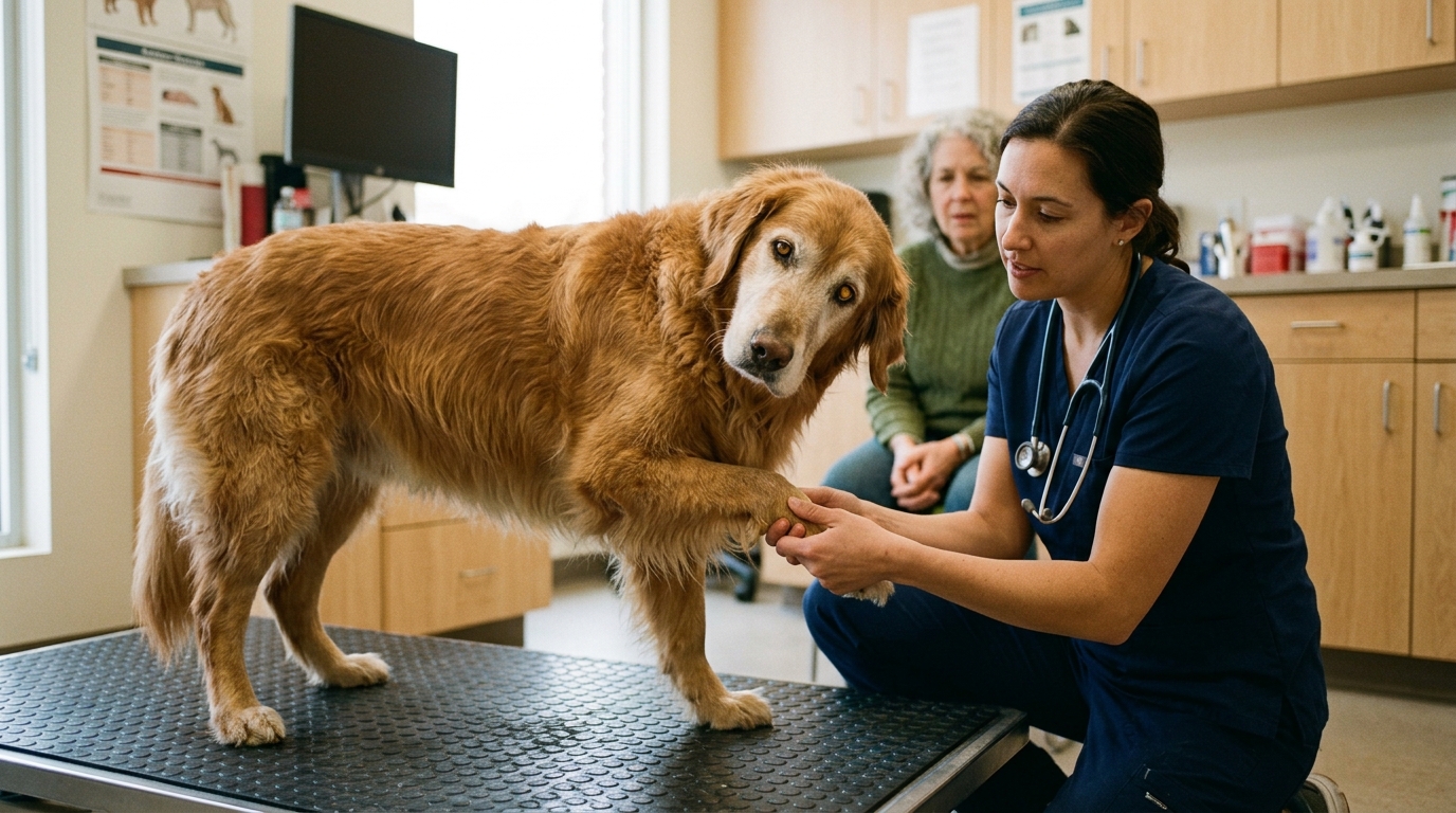
Clinical Quick Answer & Immediate Stabilization Protocol
Primary Clinical Directive for Canine Peripheral Idiopathic Vestibular Syndrome & Brainstem Vestibular Localization: Differentiating self-limiting Peripheral Vestibular Disease (affecting the inner ear semicircular canals, utricle, saccule, and Cranial Nerve VIII) from serious Central Vestibular Disease (affecting the brainstem vestibular nuclei or cerebellum) hinges on four bedside neurological signs: (1) Nystagmus direction—peripheral disease causes strictly horizontal or rotary nystagmus with the fast phase beating away from the head tilt that never changes direction when the head position is altered, whereas vertical nystagmus or positional direction-changing nystagmus indicates central brainstem pathology; (2) Conscious Proprioception—peripheral patients retain normal paw knuckling reflexes once supported, whereas central patients exhibit ipsilateral proprioceptive deficits; (3) Mentation—peripheral dogs are anxious/nauseated but alert, whereas central lesions cause stupor or obtundation; and (4) Cranial Nerves—peripheral inner-ear lesions affect only CN VIII (and occasionally adjacent CN VII facial nerve or sympathetic Horner’s fibers in middle-ear disease), never CN V, IX, X, or XII. Idiopathic peripheral disease improves markedly within 3 to 14 days with maropitant antiemetics and padded nursing care.
Vestibulo-Ocular Reflex Asymmetry, Endolymphatic Imbalance & Anatomical Localization
The vestibular system maintains posture, head position, and conjugate eye stability during movement via continuous tonic firing from hair cells inside the membranous labyrinth (three semicircular canals, utricle, and saccule) encased within the petrous temporal bone. Both the left and right vestibulocochlear nerves (Cranial Nerve VIII) send balanced baseline tonic impulses to the four paired vestibular nuclei in the medulla oblongata and pons. When unilateral pathology abruptly silences or dampens firing from one inner ear, the brainstem interprets the unopposed tonic firing from the healthy contralateral ear as continuous rapid rotation toward the healthy side.
To counteract this perceived rotation, the vestibulospinal tract reduces extensor muscle tone on the affected side while increasing extensor tone on the healthy side—causing the head to tilt, the trunk to curve, and the dog to lean, circle, or roll violently toward the side of the lesion. Simultaneously, imbalanced input to the oculomotor, trochlear, and abducens nuclei (CN III, IV, VI) drives the slow phase of pathological jerk nystagmus toward the lesion, followed by a rapid cortical corrective flick (the fast phase) away from the lesion. Because this constant visual spinning stimulates the chemoreceptor trigger zone and emetic center in the medulla, acute motion sickness, ptyalism, and vomiting are prominent during the first 48 hours.
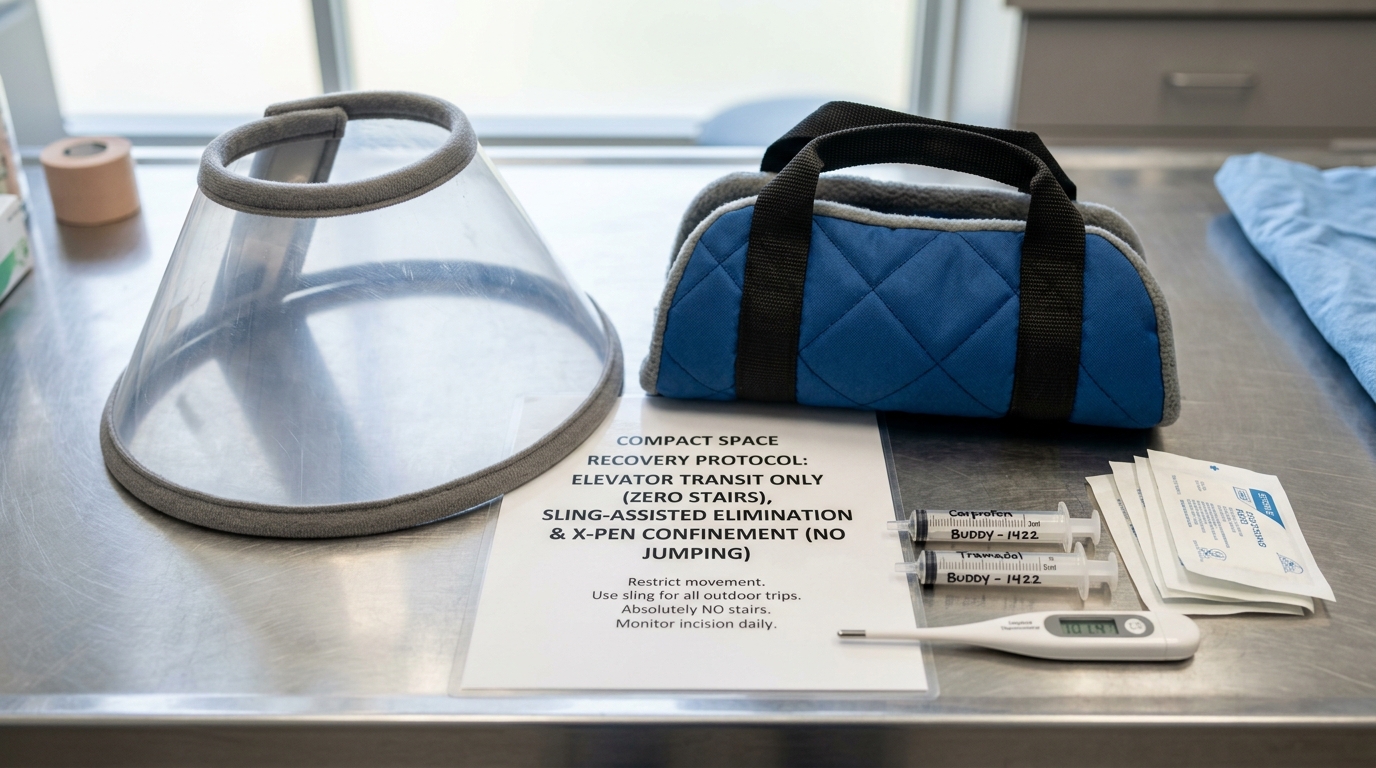
- Rule of Nystagmus Vector (Horizontal/Rotary vs. Vertical): Peripheral vestibular disease produces strictly horizontal or rotary nystagmus whose fast phase beats away from the head tilt and remains constant regardless of whether the dog is held on its back or side; spontaneous vertical nystagmus or nystagmus that flips vector with head position is pathognomonic for central brainstem/cerebellar disease.
- Otitis Media-Interna with Facial Paralysis & Horner’s Syndrome: Because the facial nerve (CN VII) and ocular sympathetic nerve fibers travel directly through the middle ear adjacent to the tympanic bulla, concurrent ipsilateral facial droop/blinking loss and Horner’s syndrome (miosis, ptosis, enophthalmos, third-eyelid protrusion) localize the lesion to the middle/inner ear rather than idiopathic senile disease.
- Paradoxical Vestibular Syndrome: Lesions of the caudal cerebellar peduncle or flocculonodular lobe produce a head tilt and circling directed away from the side of the lesion (toward the normal side), readily identified by ipsilateral conscious proprioceptive paw-knuckling deficits and hypermetria.
- 72-Hour Brainstem Compensation Window: In idiopathic geriatric peripheral vestibular syndrome, cerebellar inhibition re-balances resting activity between the left and right brainstem vestibular nuclei within 48 to 72 hours, halting spontaneous nystagmus first, restoring ambulation by days 5 to 7, and achieving near-complete recovery within 2 to 3 weeks (occasionally leaving a mild residual 5-to-10-degree head tilt).
Emergency Triage Matrix: Clinical Presentation & Required Response
| Triage Level | Clinical Signs & Presentation | Immediate Action Protocol |
|---|---|---|
| Tier 1: Immediate Emergency (0–2 Hours) | Peracute head tilt with violent rolling, inability to stand, vertical or direction-changing nystagmus, delayed paw knuckling (proprioceptive deficits), dull/stuporous mentation, or concurrent generalized seizures. | Transport immediately on a padded board or blanket to a 24-hour veterinary hospital or neurology center for central vs. peripheral localization, blood pressure check, and maropitant/ondansetron antiemetic stabilization. |
| Tier 2: Urgent Same-Day Evaluation (2–6 Hours) | Sudden onset of head tilt, horizontal/rotary darting eyes (nystagmus), wide-based drunken stumbling (ataxia), drooling, or single vomiting episode in an alert senior dog with normal paw placement. | Schedule a same-day veterinary examination including deep otoscopy, cranial nerve exam, blood pressure, thyroid T4/TSH panel, and injectable maropitant/meclizine nausea control. |
| Tier 3: 14-Day Home Vestibular Nursing & Rehabilitation | Diagnosed idiopathic peripheral vestibular dog beyond 72 hours showing resolving nystagmus, returning appetite, and steady improvement in assisted walking. | Use a padded chest-and-pelvic harness sling for outdoor bathroom trips, elevate food and water bowls to shoulder height, keep floors covered with non-slip mats, and block all stairs. |
Differential Rule-Outs (Otitis Media, Hypothyroidism, Infarction), Maropitant Antiemesis & Nursing Care
Before assigning a diagnosis of Idiopathic Geriatric Vestibular Syndrome (a diagnosis of exclusion), clinicians perform a targeted minimum database to eliminate treatable etiologies of peripheral and central vestibular dysfunction. Deep video otoscopy (and bulla radiography or CT when indicated) rules out ascending bacterial Otitis Media-Interna, nasopharyngeal polyps, or ceruminous adenocarcinoma. Systemic blood pressure measurement and complete blood count/biochemistry/urinalysis screen for hypertensive or thromboembolic cerebellar ischemic infarction (linked to protein-losing nephropathy, Cushing’s disease, or pheochromocytoma). A complete thyroid profile (total T4, free T4 by equilibrium dialysis, and cTSH) rules out hypothyroid myxedema neuropathy of CN VIII, while medication history excludes iatrogenic ototoxicity from high-dose systemic metronidazole exposure or topical chlorhexidine/aminoglycoside ear drops applied across a ruptured tympanum.
Once idiopathic peripheral vestibular syndrome is confirmed, corticosteroids and empirical antibiotics provide zero clinical benefit and are avoided. Medical therapy focuses on extinguishing debilitating vestibular nausea and emesis using the neurokinin-1 (NK-1) receptor antagonist Maropitant (Cerenia)—which blocks Substance P in the brainstem emetic center without sedating central cerebellar vestibular compensation—occasionally paired with ondansetron or meclizine, plus intravenous or subcutaneous isotonic fluids during the initial 48-hour anorexic phase.
- Maropitant Over Heavy Phenothiazine Sedation: Heavy sedatives (such as acepromazine) depress central nervous system plasticity and delay cerebellar re-calibration of the vestibular nuclei; maropitant controls severe nausea while allowing active neuro-adaptation.
- Propentofylline & Physical Proprioceptive Input: Once vomiting subsides at 48 to 72 hours, gently encouraging the dog to stand and take supported steps inside a padded harness sling stimulates visual and somatosensory proprioceptive pathways that accelerate vestibular compensation.
- Preventing Decubital Ulcers & Corneal Abrasions: Because rolling and recumbent dogs press the downwards-facing eye (on the side of the head tilt) into bedding, apply sterile artificial tear lubricating ointment every 6 hours to the dependent eye and rotate the dog on thick orthopedic foam pads every 4 hours.
- Hand-Feeding & Elevated Bowl Positioning: Lowering the head to a floor-level bowl triggers acute positional vertigo and rolling; hold water and high-aroma food bowls at chest level while steadying the dog’s shoulders against a padded wall.
Strict Rule S1 Clinical Red Flags: Canine Peripheral Idiopathic Vestibular Syndrome & Brainstem Vestibular Localization
Critical safety boundaries for Canine (Canis lupus familiaris — Senior & Geriatric Dogs Mean Age 12–13 Years) caregivers:
- Zero Ear Drops or Flushing Before Eardrum Inspection: Never squirt ear cleaners, hydrogen peroxide, vinegar, or leftover antibiotic ear drops into the ear of a dog with a head tilt; if the tympanic membrane is perforated, these fluids enter the inner ear and cause permanent deafness and severe vestibular destruction.
- Zero Premature Euthanasia in the First 72 Hours: Peripheral idiopathic vestibular disease looks alarming on Day 1, but over 90% of affected senior dogs show dramatic reduction in nystagmus within 72 hours and regain independent walking within 7 to 14 days.
Clinical Preparation & Biosecurity Action Checklist
- Film a 30-second close-up video of your dog’s eyes showing the exact direction of the eye flicking (horizontal side-to-side vs. vertical up-and-down) to show your veterinarian.
- Block off all stairs, decks, and swimming pools immediately, and clear coffee tables or sharp furniture edges around your dog’s resting mat.
- Use a rolled bath towel under the belly—or a commercial padded full-body mobility harness—to support your dog’s weight during outdoor elimination trips.
- Hold food and water bowls at chest level and hand-feed small meatballs of wet food once veterinary antiemetic injection (maropitant) has controlled nausea.
Related Clinical Guides & Triage Protocols
Peer-Reviewed Clinical References & Evidence Grounding
- Clinical features, diagnostic findings, and outcome of dogs with peripheral and central vestibular disease: 188 cases (opens in new tab) (Journal of Veterinary Internal Medicine (2020) — Peer-reviewed clinical neurology study by Bongartz et al. indexed on NCBI PubMed (PMID: 32315389).)
- Vestibular disease in dogs and cats: neuroanatomical localization and differential diagnosis (opens in new tab) (Veterinary Clinics of North America: Small Animal Practice (2014) — Peer-reviewed neurology reference by Rossmeisl indexed on NCBI PubMed (PMID: 25483191).)
- 2023 AAHA Senior Care Guidelines for Dogs and Cats — Geriatric Neurological and Mobility Management (opens in new tab) (Journal of the American Animal Hospital Association (2023) — AAHA Consensus Guidelines indexed on NCBI PubMed (PMID: 36695816).)
- Merck Veterinary Manual: Vestibular Disease in Dogs and Cats (opens in new tab) (Verified veterinary clinical reference on horizontal vs vertical nystagmus localization, otitis media-interna, and idiopathic geriatric syndrome.)
Source transparency
Reference mix for this guide
Source labels classify the institutional category of the references cited above.