This guide is general education for pet owners. If your pet has urgent symptoms, possible poisoning, injury, breathing trouble, collapse, severe pain, or sudden decline, contact a licensed veterinarian or local emergency service.
Dosages must be calculated by a licensed veterinarian based on species, weight, and bloodwork. Never administer human medications or self-adjust prescription doses for pets.
Editorial standard
How this guide was prepared
In small-animal emergency medicine, the adage “never let the sun set on a red, squinting, or cloudy eye” is a strict physiological imperative. A dog or cat presenting with sudden blepharospasm (squinting), epiphora (tearing), episcleral blood-vessel engorgement, blue corneal edema, or unequal pupil sizes (anisocoria) cannot be safely triaged by visual inspection at home because four radically different conditions—corneal ulceration, acute primary glaucoma, anterior uveitis, and anterior lens luxation—all produce a painful “red, cloudy eye.” Misapplying a leftover steroid eye drop to an ulcerated cornea activates collagenase enzymes that melt the eye open within 24 hours, while waiting overnight with acute glaucoma permanently crushes the optic nerve head.
Clinical Quick Answer & Immediate Stabilization Protocol
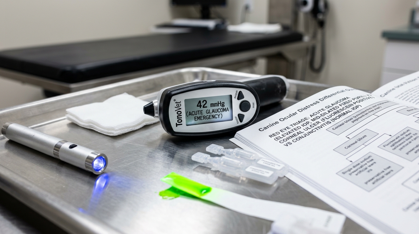
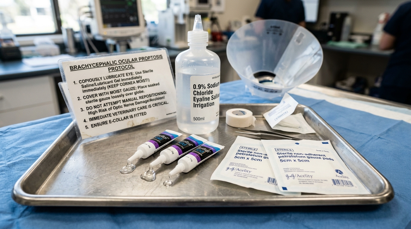
Primary Clinical Directive for Melting Corneal Ulcer / Descemetocele, Acute Congestive Glaucoma & Traumatic Globe Proptosis: Every acute eye symptom works on a 1-to-6-hour vision-saving clock: (1) Traumatic Globe Proptosis (eyeball displaced forward of the eyelids after trauma or neck scruffing in a brachycephalic dog) or Perforated/Melting Corneal Ulcer requires emergency veterinary arrival within 30 to 60 minutes while keeping the exposed cornea moist with sterile saline or artificial tear gel (never push the eyeball back in yourself); (2) Acute Angle-Closure Glaucoma (cloudy blue cornea, dilated fixed pupil, cobweb-red sclera, squinting, lethargy) requires emergency intraocular pressure lowering (mannitol/latanoprost) within 2 to 4 hours to save retinal ganglion cells; and (3) Any New Squinting or Rubbing requires an E-collar immediately and a same-day veterinary fluorescein stain, Schirmer tear test, and tonometry.
Stromal Collagenase Melting, Retinal Ganglion Pressure Axotomy & Brachycephalic Lagophthalmos
The transparent canine and feline cornea measures less than 0.6 to 0.8 millimeters thick and consists of four primary layers: the hydrophobic outer stratified squamous epithelium, the hydrophilic collagenous stroma (comprising 90% of thickness), Descemet’s basement membrane, and the single-cell inner endothelium. When a cat claw, thorny bush, entropion eyelash, or desiccated brachycephalic tear film breaches the outer epithelium, fluorescein dye binds to the exposed hydrophilic stroma, glowing neon green under cobalt-blue light. If opportunistic bacteria (particularly Pseudomonas aeruginosa or beta-hemolytic Streptococci) or topical steroid drops contaminate the stromal defect, bacterial proteases and host matrix metalloproteinases (MMP-2 and MMP-9) liquefy the collagen lamellae (“keratomalacia” or melting ulcer) within 12 to 24 hours. Once the stroma dissolves down to the 10-micrometer-thick Descemet’s membrane (a “descemetocele,” which curiously does NOT retain fluorescein stain in its clear center, tricking untrained observers into thinking the ulcer healed!), even a minor bark or collar pull ruptures the globe.
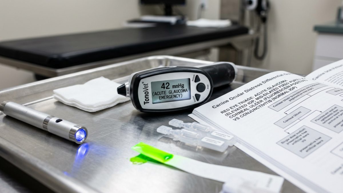
In acute primary angle-closure glaucoma (common in Cocker Spaniels, Basset Hounds, Beagles, Chow Chows, Huskies, and Shiba Inus) or secondary glaucoma from anterior lens luxation (Terriers), obstruction of aqueous humor outflow through the iridocorneal pectinate ligament drives intraocular pressure (IOP) from a normal 10–22 mmHg up to 50–80+ mmHg within hours. This immense hydrostatic pressure compresses the optic nerve axons as they exit the lamina cribrosa, halting axoplasmic transport and causing irreversible ischemic blindness within 4 to 24 hours.
- The Descemetocele “Clear-Center” Trap: Because Descemet’s membrane is hydrophobic, a deep crater ulcer that has eaten through 95% of the cornea shows a green fluorescein ring with a dark clear bottom—requiring emergency conjunctival graft surgery by a veterinary ophthalmologist rather than routine drops.
- Glaucoma vs. Uveitis Pupil & IOP Differential: Both glaucoma and anterior uveitis cause a painful red, cloudy eye, but uveitis presents with a constricted (miotic) pupil and LOW intraocular pressure (<10 mmHg), whereas acute glaucoma presents with a dilated (mydriatic), unreactive pupil and HIGH pressure (>30–50 mmHg).
- Proptosis Restraint Trigger in Brachycephalics: Pugs, Shih Tzus, Pekingese, and French Bulldogs have shallow bony orbits and large palpebral fissures; excessive neck-skin scruffing or tight collar tension can pop the globe forward of the eyelids (proptosis), trapping the eyelids behind the equator of the eye.
- Melting Ulcer Anticollagenase Protocol: Emergency treatment of a gelatinous melting corneal ulcer requires hourly topical autologous serum (containing alpha-2 macroglobulin anticollagenases), topical acetylcysteine/EDTA, systemic doxycycline, and intensive broad-spectrum fluoroquinolone/cefazolin drops.
Emergency Triage Matrix: Clinical Presentation & Required Response
| Triage Level | Clinical Signs & Presentation | Immediate Action Protocol |
|---|---|---|
| Tier 1: Immediate Ophthalmic Emergency (0–2 Hours) | Eyeball bulging forward past eyelids (proptosis), clear dark crater or gelatinous dripping/jelly appearance on cornea, black/brown iris tissue protruding from eye, or acute blue cloudy eye with a wide dilated pupil and screaming/lethargy. | Put a rigid plastic E-collar on the pet immediately, replace any neck collar with a chest harness (neck pressure spikes IOP and ruptures descemetoceles), apply sterile lubricating eye gel (only if non-perforated proptosis) without pressing on the eyeball, and drive directly to an emergency vet or veterinary ophthalmologist. |
| Tier 2: Urgent Same-Day Evaluation (2–8 Hours) | Sudden squinting (blepharospasm) of one or both eyes, pawing or rubbing face on carpet, thick yellow-green ocular discharge, red bloodshot sclera, or third eyelid (nictitating membrane) elevated across the eye. | Fit an E-collar immediately, keep the pet in a dimly lit room, do not instill any home or old prescription drops, and obtain a same-day veterinary examination for Schirmer tear testing, fluorescein staining, and tonometry. |
| Tier 3: Post-Diagnosis Drop Administration & Recheck Compliance | Diagnosed superficial uncomplicated corneal abrasion on prescribed topical antibiotic drops and oral analgesia. | Space multiple different eye drops at least 5 to 10 minutes apart (liquids before ointments), keep the E-collar on 24/7 until the 72-hour re-stain confirms 100% epithelial closure, and watch for any sudden worsening of squinting. |
The Mandatory Three-Step Ocular Workup (STT, Fluorescein, Tonometry) & Proper Drop Sequencing
Every red or painful eye presented to a veterinary clinic must undergo the three-step ophthalmic diagnostic triad in a strict sequential order: Step 1 is the Schirmer Tear Test (STT-1), performed BEFORE any liquid drops or local anesthetic touch the eye, to diagnose keratoconjunctivitis sicca (“dry eye,” <15 mm/min in dogs). Step 2 is Sodium Fluorescein Staining, flushed with sterile saline and examined under cobalt-blue illumination to map epithelial defects, dendritic feline herpesvirus ulcers, or deep stromal craters. Step 3 is Rebound or Applanation Tonometry (TonoVet / Tono-Pen) to measure exact intraocular pressure in millimeters of mercury—taking utmost care never to press on the jugular veins or eyelids during measurement, which falsely spikes IOP readings by 10 to 20 mmHg.
When administering multiple prescribed ophthalmic medications at home, caregivers must wait 5 to 10 minutes between different bottles. Because the canine and feline conjunctival cul-de-sac holds only 1 drop (approx. 30–50 microliters), squirting a second medication 30 seconds after the first washes 90 percent of the first drug straight down the nasolacrimal duct. Always apply watery solutions first, serum/suspensions second, and thick viscous gels or ointments last.
- Topical Atropine Precaution in Dogs & Cats: Veterinary ophthalmologists often prescribe a single dose of topical 1% atropine to relieve painful ciliary muscle spasm (cycloplegia) in corneal ulcers, but atropine is strictly contraindicated in glaucoma and KCS dry eye—and because atropine tastes intensely bitter when it drains via the tear duct into the mouth, cats frequently foam and drool profusely for 10 minutes after a drop (not a toxic seizure!).
- Never Use a Soft Inflatable Donut Collar for Eye Injuries: Soft inflatable neck donuts and flexible fabric cones allow dogs and cats to rub their cornea against sofa corners, table legs, and their own front dewclaws; only a rigid plastic Elizabethan cone extending 2 inches past the nose tip protects the cornea.
- Topical Proparacaine Is Diagnostic Only—Never a Treatment: Local anesthetic drops (proparacaine/tetracaine) numb the cornea for 15 minutes during exam, but repeated use is directly toxic to corneal epithelial migration and causes non-healing perforation.
- Prophylactic Fellow-Eye Glaucoma Protection: Primary angle-closure glaucoma is a bilateral genetic disease; when one eye suffers an acute glaucoma attack, starting prophylactic pressure-lowering drops (dorzolamide/timolol or demecarium) in the healthy “normal” eye delays second-eye blindness by more than 20 months.
Strict Rule S1 Clinical Red Flags: Melting Corneal Ulcer / Descemetocele, Acute Congestive Glaucoma & Traumatic Globe Proptosis
Critical safety boundaries for Canine & Feline (Canis lupus familiaris & Felis catus — Brachycephalic, Terrier & All Breeds) caregivers:
- Zero Leftover Steroid Eye Drops (Dexamethasone / Prednisolone / Neo-Poly-Dex) in Unstained Eyes: Never put a leftover anti-inflammatory eye drop into a squinting pet’s eye; if an invisible corneal scratch is present, topical steroids trigger catastrophic collagenase melting and eye loss within 24–48 hours.
- Zero Manual Attempt to Push a Proptosed Eyeball Back Into the Socket at Home: Never press on a displaced eyeball with fingers or gauze; manual compression without lateral canthotomy under general anesthesia ruptures the globe and avulses the optic nerve.
Clinical Preparation & Biosecurity Action Checklist
- Keep a properly sized rigid plastic Elizabethan cone collar on hand at home and put it on your pet the very first minute you notice squinting or eye pawing.
- Switch any dog with corneal disease, glaucoma, or brachycephalic anatomy permanently from a neck collar to a Y-front chest harness to prevent jugular pressure spikes.
- Wait a full 5 to 10 minutes between different prescribed eye drops (applying thin liquids first and thick gels/ointments last) and never touch the dropper tip to the eyelashes.
- Discard all open prescription eye drop bottles once a treatment course finishes so no caregiver accidentally reuses a steroid drop on a future corneal ulcer.
Related Clinical Guides & Triage Protocols
Peer-Reviewed Clinical References & Evidence Grounding
- Ophthalmic emergencies in small animals: diagnosis and management of acute glaucoma, corneal perforation, and traumatic proptosis (opens in new tab) (Veterinary Clinics of North America: Small Animal Practice (2017) — Peer-reviewed ophthalmology emergency review indexed on NCBI PubMed (PMID: 28941312).)
- Effect of prophylactic topical antiglaucoma therapy on the time to onset of primary glaucoma in the second eye of dogs (opens in new tab) (Journal of the American Veterinary Medical Association (2001) — Landmark clinical study by Miller et al. indexed on NCBI PubMed (PMID: 11720273).)
- American College of Veterinary Ophthalmologists (ACVO) Clinical Standards on Corneal Ulceration, Tonometry, and Glaucoma Triage (opens in new tab) (Board-certified veterinary ophthalmology specialty guidelines.)
- Merck Veterinary Manual: Ophthalmic Emergencies in Small Animals (opens in new tab) (Verified veterinary clinical reference on melting corneal ulcers, descemetoceles, acute glaucoma, and proptosis.)
Source transparency
Reference mix for this guide
Source labels classify the institutional category of the references cited above.