This guide is general education for pet owners. If your pet has urgent symptoms, possible poisoning, injury, breathing trouble, collapse, severe pain, or sudden decline, contact a licensed veterinarian or local emergency service.
Dosages must be calculated by a licensed veterinarian based on species, weight, and bloodwork. Never administer human medications or self-adjust prescription doses for pets.
Editorial standard
How this guide was prepared
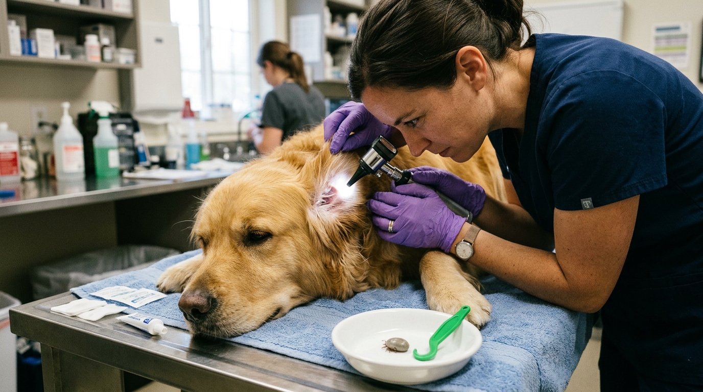
Finding an engorged hard tick (Ixodes scapularis/pacificus, Dermacentor variabilis/andersoni, Amblyomma americanum, Rhipicephalus sanguineus, or Ixodes holocyclus) anchored into a dog or cat’s skin requires rapid, mechanically precise extraction—yet traditional folk remedies such as coating the tick in petroleum jelly, touching it with a hot match, dabbing rubbing alcohol or nail polish, or twisting the bloated abdomen with fingers actually trigger the exact pathogen transmission the owner is trying to prevent. Furthermore, beyond transmitting bacterial and protozoal pathogens (Borrelia burgdorferi, Ehrlichia canis/ewingii, Anaplasma phagocytophilum, Rickettsia rickettsii, Babesia, and feline Cytauxzoon felis), a single gravid female tick hidden inside the ear fold or between the toes can secrete a salivary neurotoxin that blocks acetylcholine release at the neuromuscular junction, producing rapidly ascending flaccid paralysis and respiratory arrest (“tick paralysis”).
Clinical Quick Answer & Immediate Stabilization Protocol
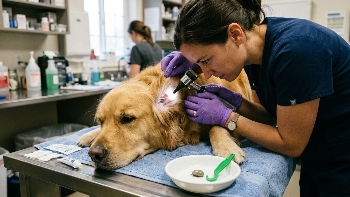
Primary Clinical Directive for Ixodid Tick Attachment, Ascending Tick Paralysis & Tick-Borne Rickettsial/Spirochetal Triage: Extract every attached tick immediately using fine-point tweezers or a slotted tick-removal hook (Tick Twister / O’Tom): part the fur, grasp the tick’s hard head/mouthparts (capitulum) as close to the skin surface as possible—never squeezing the swollen gray/brown belly—and pull straight upward with steady, even pressure until the skin tents and the cement plug releases. Drop the tick into a small sealed jar or zip-bag with a splash of rubbing alcohol (saved with the date written on the bag for identification if symptoms develop), dab the bite site with chlorhexidine or povidone-iodine, and calendar a veterinary 4Dx + CBC blood test for 4 to 6 weeks post-bite (or seek immediate emergency veterinary care if hindlimb wobbliness, fever, lameness, or anorexia appears sooner).
Salivary Transmission Time-Windows, Presynaptic Tick Neurotoxin & Lyme Nephritis Screening
Tick-borne pathogens reside in different anatomical compartments within the unfed tick vector, dictating how many hours of blood-meal attachment must elapse before transmission occurs. Spirochetes of Borrelia burgdorferi (the agent of Lyme disease, vectored by Ixodes scapularis and Ixodes pacificus) lie dormant inside the tick’s midgut; upon sensing warm host blood, the spirochetes down-regulate outer surface protein A (OspA), up-regulate OspC, migrate through the hemolymph to the salivary glands, and enter the dog roughly 24 to 48 hours after attachment. In contrast, rickettsial pathogens already primed inside the salivary glands—including Ehrlichia canis (transmitted within 3 to 6 hours by Rhipicephalus sanguineus), Anaplasma phagocytophilum (12 to 24 hours), Rickettsia rickettsii (5 to 20 hours), and feline Cytauxzoon felis (36 hours by Amblyomma americanum)—infect the host far earlier, explaining why fast-kill oral isoxazolines and prompt daily physical tick checks must operate together.
In Tick Paralysis, an engorging adult female tick (after 4 to 7 days of feeding) secretes neurotoxic salivary holocyclotoxins or neurotoxins that inhibit presynaptic voltage-gated calcium influx at the motor nerve terminal, preventing acetylcholine vesicle exocytosis. The dog or cat develops symmetrical, afebrile, ascending lower motor neuron (LMN) flaccid paralysis starting in the hindlimbs (ataxia progressing to paraplegia within 12–24 hours), advancing to the forelimbs (quadriplegia with absent patellar/withdrawal reflexes while pain sensation and tail wag often remain preserved initially), dysphonia (altered bark), megaesophagus, and fatal diaphragmatic respiratory failure. In North American Dermacentor tick paralysis, simply finding and plucking the single culprit female tick typically produces dramatic neurological recovery within 24 to 72 hours (whereas Australian Ixodes holocyclus paralysis continues to worsen for 24–48 hours after tick removal and requires specific hyperimmune Tick Antiserum ICU therapy).
- What If the Tick’s Mouthpart (Hypostome) Breaks Off in the Skin? Leave It Alone: If a tiny black speck of the barbed hypostome or cement cone snaps off inside the dermis during removal, do NOT dig into your dog’s skin with a sewing needle or tweezers (which causes deep staphylococcal cellulitis); the pet’s epidermis will harmlessly slough the sterile chitin shard out like a tiny splinter over 5 to 7 days.
- The Normal “Tick Granuloma” Bump: A firm, pea-sized, non-painful skin nodule at the tick bite site is a normal localized reaction to tick salivary cement proteins and resolves over 2 to 4 weeks without antibiotics.
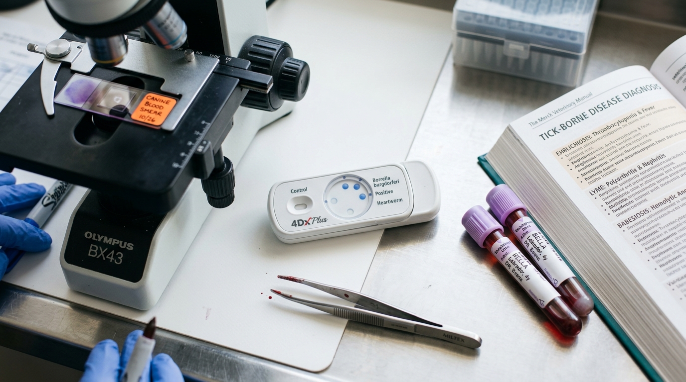
- Why Testing a Dog the Day After a Tick Bite Yields a False Negative: Point-of-care 4Dx Plus SNAP tests detect host antibodies (such as the C6 peptide antibody for Borrelia), which require 3 to 4 weeks for the dog’s immune system to produce after a bite; testing 24 hours after pulling a tick cannot detect the new exposure.
- Lyme Nephritis & Urine Protein Screening (UPC): While 90% of Borrelia-seropositive dogs remain subclinical, Golden Retrievers, Labradors, and Shetland Sheepdogs face a high risk of fatal immune-complex Lyme glomerulonephritis; ACVIM guidelines mandate urinalysis / urine protein-to-creatinine ratio (UPC) screening for every Lyme C6-positive dog.
Emergency Triage Matrix: Clinical Presentation & Required Response
| Triage Level | Clinical Signs & Presentation | Immediate Action Protocol |
|---|---|---|
| Tier 1: Ascending Hindlimb Paralysis, Petechial Bleeding or Feline High Fever (0–2 Hours) | Dog or cat develops wobbly hind legs progressing to inability to stand (tick paralysis), pinpoint red gum spots / nosebleeds (acute Ehrlichia/Rickettsia thrombocytopenia), or a cat with outdoor access develops sudden high fever (>104°F), jaundice, and vocal lethargy (Cytauxzoonosis). | Transport immediately to an emergency veterinary hospital; perform a systematic millimeter-by-millimeter skin search (including inside ear canals, between toes, under the collar, and perineum) to remove all ticks, support ventilation, and initiate emergency rickettsial/protozoal therapy. |
| Tier 2: Shifting-Leg Lameness, Swollen Joints & Fever 2–20 Weeks Post-Tick (12–24 Hours) | Dog suddenly limps on one front or hind leg with a warm swollen carpal/stifle joint, fever (103°F–104.5°F), swollen lymph nodes, and lethargy weeks or months after tick season. | Schedule a same-day or next-day veterinary exam for a 4Dx Plus serology panel, CBC (checking platelet count for Anaplasma/Ehrlichia thrombocytopenia), urinalysis for proteinuria, and a 28-day course of oral doxycycline. |
| Tier 3: Freshly Removed Tick on an Asymptomatic, Bright Pet | Tick removed cleanly with forceps; small 3-mm pink bump at the attachment site; pet has normal appetite, normal gait, and normal temperature. | Clean the bite site with chlorhexidine antiseptic, save the tick in a dated alcohol vial for 30 days, verify monthly isoxazoline preventative compliance, and test on 4Dx serology 4–6 weeks post-bite. |
Precision Forceps Extraction, The Eight-Hotspot Post-Walk Body Search & ACVIM Lyme Consensus
After every walk near brush, tall grass, leaf litter, or stone walls (including autumn and mild winter thaws above 39°F / 4°C when adult blacklegged Ixodes ticks actively quest), perform a 2-minute tactile “Eight-Hotspot Tick Check.” Because unfed nymphal ticks are the size of a poppy seed (1.5 mm), visual scanning misses them; run your fingertips slowly against the grain of the fur across the eight anatomical warm zones where ticks migrate before biting: (1) inside and behind the ear flaps (and Henry’s pocket), (2) directly beneath the neck collar or harness straps, (3) around the eyelids and chin whiskers, (4) in the axillary armpits, (5) between all four paw pads and interdigital webs, (6) in the inguinal groin fold, (7) under the base of the tail/perineum, and (8) along the dorsal midline.
In accordance with the ACVIM Consensus Statement on Lyme Borreliosis in Dogs and Cats, asymptomatic dogs that test C6-antibody positive on a routine screening 4Dx test should first undergo a complete urinalysis (and Urine Protein:Creatinine ratio if proteinuria is detected) plus a CBC to check for co-infection with Anaplasma phagocytophilum platelets. Dogs with clinical polyarthritis (shifting-leg lameness and fever) or significant proteinuria receive a full 4-week (28-day) course of doxycycline.
- Fine-Point Tweezers vs. Slotted Tick Hooks: Avoid broad, flat eyebrow tweezers (which crush the bloated abdomen of an engorged tick); keep a dedicated pair of needle-point curved forceps or a V-slotted tick pry tool (which slides beneath the mouthparts without compressing the tick’s midgut) on your leash rack.
- Isoxazoline Systemic Speed-of-Kill: Modern veterinary oral/topical isoxazolines (fluralaner, afoxolaner, sarolaner, lotilaner) circulate in systemic tissue fluids and kill attaching ticks within 8 to 12 hours of bite onset—blocking Borrelia transmission before the 24–48 hour midgut migration window completes.
- Shaving Focal Fur Mats in Paralysis Searches: When a long-haired dog (Australian Shepherd, Golden Retriever, Newfoundland) presents with acute lower motor neuron paralysis, veterinary teams frequently find 1 to 3 additional hidden ticks inside the ear canal, lip folds, or matted tail base even after the owner removes one visible tick; always search the entire dog twice.
- Feline Cytauxzoonosis Prevention: For cats in the South-Central and Southeastern United States, even a single Lone Star tick (Amblyomma americanum) walking onto a cat from a dog’s coat or screen porch can transmit fatal Cytauxzoon felis; keep all cats on year-round feline-labeled tick prevention (such as fluralaner/sarolaner topical).
Strict Rule S1 Clinical Red Flags: Ixodid Tick Attachment, Ascending Tick Paralysis & Tick-Borne Rickettsial/Spirochetal Triage
Critical safety boundaries for Canine & Feline (Canis lupus familiaris & Felis catus — All Outdoor & Suburban Pets) caregivers:
- Zero Matches, Petroleum Jelly, Nail Polish, or Alcohol on Attached Live Ticks: Never attempt to “back a tick out” with heat, grease, or chemicals; noxious stimuli cause immediate tick salivary/midgut regurgitation into the dermal capillary bed.
- Zero Canine Permethrin Spot-On Products Applied to Cats (Or Cats Grooming Newly Treated Dogs): Never apply a dog-labeled permethrin, deltamethrin, or cyphenothrin tick spot-on to a cat, and separate cats from permethrin-treated dogs until the dog’s fur is 100% dry; feline hepatic glucuronidation deficiency causes severe permethrin tremor seizures and death.
Clinical Preparation & Biosecurity Action Checklist
- Mount a pair of fine-point tick forceps or a slotted Tick Twister hook and a small dated specimen vial right beside your dog’s leashes at the front door.
- Perform the 2-minute fingertip Eight-Hotspot Tick Check (ears, under collar, eyelids, armpits, between toes, groin, tail base) immediately after every trail or park walk.
- Maintain 12-month continuous veterinary-prescribed tick prevention for both dogs and cats, as adult Ixodes deer ticks quest actively on any winter day above 39°F (4°C).
- Mark your calendar for 35 days (5 weeks) after any engorged tick removal to request a veterinary 4Dx Plus antibody test, platelet count, and urinalysis.
Related Clinical Guides & Triage Protocols
Peer-Reviewed Clinical References & Evidence Grounding
- ACVIM consensus update on Lyme borreliosis in dogs and cats: C6 serology, proteinuria screening, and antimicrobial stewardship (opens in new tab) (Journal of Veterinary Internal Medicine (2018) — ACVIM Consensus Statement by Littman et al. indexed on NCBI PubMed (PMID: 29566442).)
- Tick transmission times for Borrelia burgdorferi, Anaplasma phagocytophilum, and Ehrlichia species and speed-of-kill prevention kinetics (opens in new tab) (Parasites & Vectors (2018) — Peer-reviewed parasitology review indexed on NCBI PubMed (PMID: 29784368).)
- Companion Animal Parasite Council (CAPC) Clinical Guidelines on Tick Control, Mechanical Extraction, and Tick Paralysis (opens in new tab) (Veterinary parasitology consensus standards on year-round vector prevention and removal.)
- Merck Veterinary Manual: Tick Paralysis in Animals — Presynaptic Acetylcholine Blockade and Ascending LMN Recovery (opens in new tab) (Verified veterinary neurology reference on Dermacentor and Ixodes tick paralysis.)
Source transparency
Reference mix for this guide
Source labels classify the institutional category of the references cited above.