This guide is general education for pet owners. If your pet has urgent symptoms, possible poisoning, injury, breathing trouble, collapse, severe pain, or sudden decline, contact a licensed veterinarian or local emergency service.
Dosages must be calculated by a licensed veterinarian based on species, weight, and bloodwork. Never administer human medications or self-adjust prescription doses for pets.
Editorial standard
How this guide was prepared
When a dog or cat begins shaking its head violently and scratching at one or both ears—often developing a sudden, warm, pillow-like fluid swelling of the ear flap (an aural or auricular hematoma) overnight—the swollen ear flap is only the mechanical symptom; the underlying driver resides deeper inside the L-shaped external auditory canal. Over 90 percent of aural hematomas are triggered by intense pruritus or pain from acute otitis externa (caused by atopic allergy flares, Malassezia yeast, Staphylococcus pseudintermedius, Pseudomonas aeruginosa, ear mites Otodectes cynotis, or a migrating plant awn/foxtail). Simply draining the swollen ear flap with a needle at home or squirting home vinegar/peroxide mixtures into an unexamined ear canal guarantees hematoma recurrence and risks irreversible inner-ear deafness if the eardrum is ruptured.
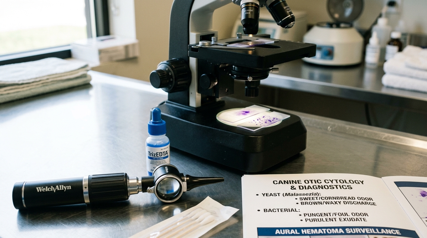
Clinical Quick Answer & Immediate Stabilization Protocol
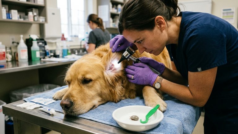
Primary Clinical Directive for Auricular (Aural) Pinna Hematoma, Acute Otitis Externa & Tympanic Membrane Rupture Risk: If your pet’s ear flap swells up like a warm water balloon (aural hematoma) or your pet cries, tilts its head, and shakes violently, schedule a veterinary examination within 24 to 48 hours (or same-day emergency triage if a balance loss, facial droop, or rolling eye movements appear). The veterinarian must perform three steps in unison: (1) Otoscopy to rule out a foreign grass awn (foxtail) and verify eardrum integrity; (2) Microscopic Ear Cytology to identify exact yeast, cocci, rods, or mites; and (3) Aural Hematoma Decompression (closed-suction cannula drainage with intralesional/systemic anti-inflammatory steroid therapy or surgical quilting sutures) so the pinna does not contract into a scarred, painful “cauliflower ear.”
Centrifugal Cartilage Shearing, L-Shaped Canal Anatomy & Ototoxicity Across Ruptured Tympana
The external ear canal of dogs and cats is not a straight horizontal tube like the human ear; it travels vertically downward along the side of the skull before making a sharp 75-to-90-degree bend inward (the horizontal canal) to meet the delicate tympanic membrane. This L-shaped anatomy traps moisture, ceruminous wax, and inflammatory exudate while preventing a cotton swab pushed from the top from doing anything other than compacting debris tightly against the eardrum like a cannon ramrod. When allergic skin inflammation (atopy or adverse food reaction) heats the ear canal lining, resident Malassezia pachydermatis yeast or opportunistic bacteria multiply rapidly, triggering intense neurogenic itching and violent head shaking.
During rapid head shaking, the tip of the ear pinna whips back and forth at high G-forces. This centrifugal shear force fractures the hyaline auricular cartilage and ruptures branches of the caudal auricular artery that pierce the cartilage plate to supply the concave inner skin. Arterial blood accumulates under pressure within the split cartilage plane, detaching the perichondrium and ballooning the ear flap into a tense, fluctuant aural hematoma. If left untreated for more than 7 to 10 days, fibrin clots organize into dense fibrous scar tissue and chondrogenesis that permanently crumples and thickens the pinna (“cauliflower ear”), obstructing airflow into the vertical canal and predisposing the pet to lifelong chronic otitis.
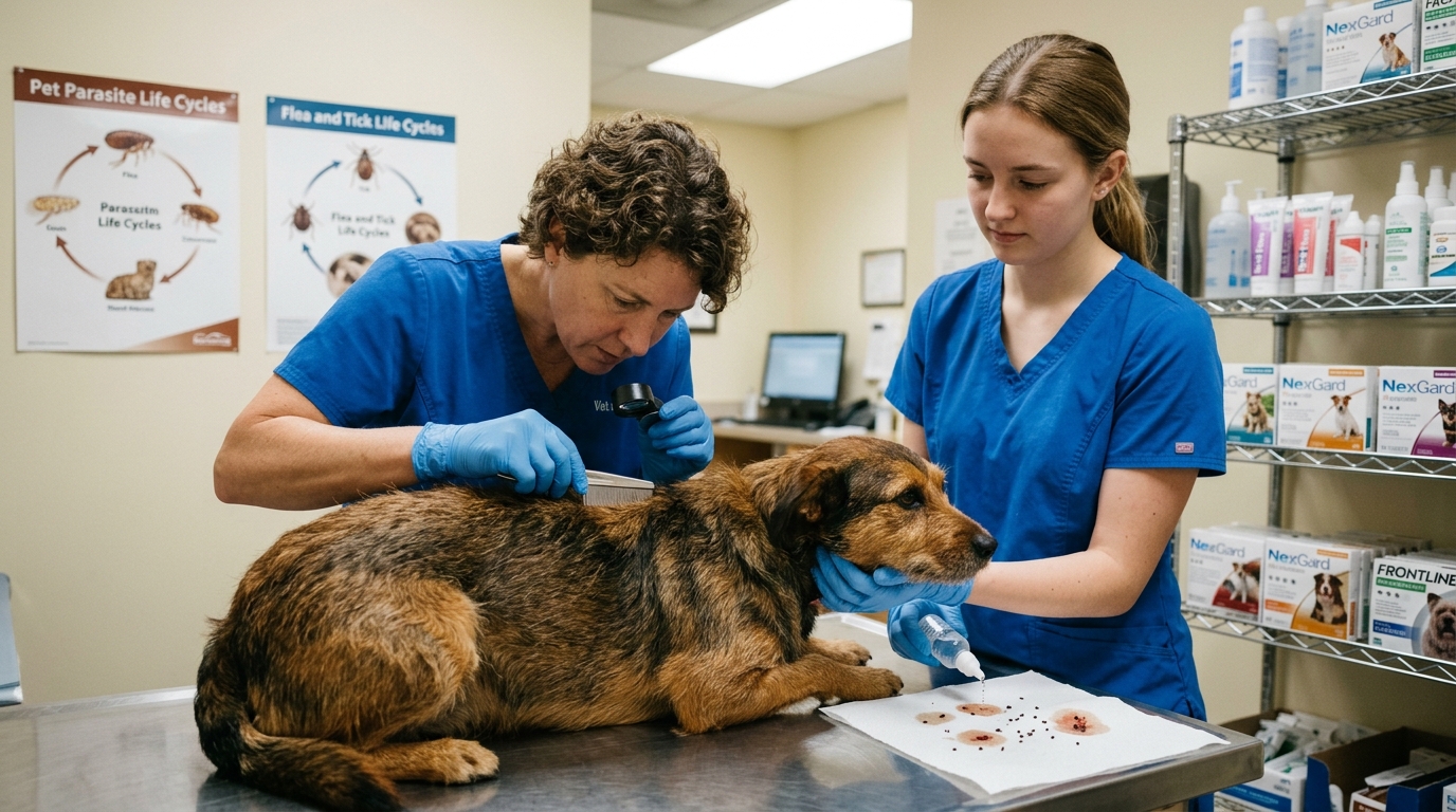
- Primary vs. Secondary Causes of Otitis (PSPP System): Yeast and bacteria are secondary opportunistic infections; unless the Primary cause (80% canine atopic dermatitis or food allergy; feline ear mites or nasopharyngeal polyps) is controlled, the ear infection and hematoma will recur within weeks.
- Pseudomonas Biofilm & Ulceration: A foul-smelling, yellow-green purulent ear discharge accompanied by extreme pain (crying or snapping when the ear base is touched) strongly suggests gram-negative Pseudomonas aeruginosa, which secretes elastases that erode the tympanic membrane in over 30% of cases.
- Otitis Media, Horner’s Syndrome & Facial Nerve Paralysis: Because the sympathetic nerve trunk and facial nerve (CN VII) travel directly past the tympanic bulla of the middle ear, extension of infection across a ruptured eardrum causes ipsilateral drooping lip/ear, inability to blink, and Horner’s triad (miosis, ptosis, enophthalmos, protruding third eyelid).
- Why Simple Needle Aspiration Fails 80%+ of the Time: Sticking a syringe needle into an aural hematoma without leaving a fenestrated drain or quilting sutures leaves a dead-space cavity that refills with blood within 24 hours.
Emergency Triage Matrix: Clinical Presentation & Required Response
| Triage Level | Clinical Signs & Presentation | Immediate Action Protocol |
|---|---|---|
| Tier 1: Acute Otitis Media/Interna or Foxtail Tympanic Penetration (0–4 Hours) | Sudden severe head tilt, loss of balance (falling or circling), involuntary rhythmic eye flicking (nystagmus), asymmetrically small pupil with raised third eyelid (Horner’s), or violent relentless pawing/screaming minutes after running through dry foxtail grass. | Do not put any liquid or cleanser into the ear canal; transport immediately for sedated video otoscopy to remove migrating grass awns before they pierce the eardrum and to initiate systemic middle-ear therapy. |
| Tier 2: Fluctuant Swollen Ear Flap (Aural Hematoma) or Purulent Painful Otitis (12–24 Hours) | Ear pinna is swollen, warm, and squishy like a fluid pillow; or ear canal is fiery red, swollen shut, malodorous, and painful on palpation with brown/yellow exudate. | Schedule a veterinary visit within 24 hours; refrain from cleaning the ear prior to the exam so cytology swabs remain diagnostic, and protect the swollen pinna from self-trauma with an E-collar or head wrap. |
| Tier 3: Maintenance Ceruminolytic Flushing in Confirmed Intact-Tympanum Ears | Pet with diagnosed atopic otitis on a veterinarian-prescribed weekly maintenance ear-cleaning and topical proactive steroid (“weekend therapy”) regimen. | Fill the canal with vet-approved pH-balanced drying cleanser, massage the base of the vertical cartilage for 30 seconds (“squish-squish” sound), let the pet shake, and wipe only the visible outer fold with a cotton ball. |
Cytology-Guided Topical Therapy, Aural Hematoma Drainage Techniques & Safe Canal Flushing
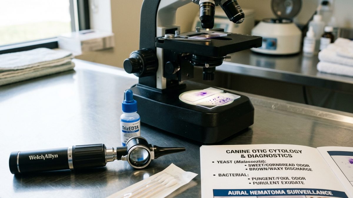
Before any ear cleanser or medication is selected, the veterinarian collects an exudate sample from the junction of the vertical and horizontal canals using a soft rayon swab, rolls it onto a glass slide, stains it with modified Wright’s stain (Diff-Quik), and examines it under 1000x oil-immersion microscopy. Finding peanut-shaped budding Malassezia yeast (>5 per high-power field) calls for topical azole/terbinafine antifungals; gram-positive cocci (Staphylococcus) respond to first-line florfenicol/mometasone or fusidic acid; whereas gram-negative rods (Pseudomonas) mandate a bacterial culture and susceptibility panel alongside Tris-EDTA biofilm-disrupting flushes.
To resolve an aural hematoma before fibrosis sets in, two primary veterinary techniques are utilized: (1) Surgical S-Incision and Thru-and-Thru Quilting Sutures under general anesthesia—incising the concave skin to evacuate fibrin clots and placing staggered vertical mattress sutures parallel to the major blood vessels to obliterate dead space while leaving the incision gap open for drainage; or (2) Minimally Invasive Teat-Cannula / Closed-Suction Drainage combined with a tapering course of anti-inflammatory corticosteroids to halt the underlying otitic head shaking.
- Never Clean the Ears the Morning of Your Vet Visit: Pouring ear cleaner into your dog’s ears two hours before a veterinary appointment washes away the surface cells and microbes needed for cytology and masks the eardrum in white foam.
- Cotton Balls Only—Zero Cotton Swabs (Q-Tips): Use soft cotton balls or gauze squares wrapped around your index finger to wipe only the visible outer bowl of the ear flap after your pet shakes out the liquid cleanser.
- How to Perform a Proper 30-Second Vertical Canal Massage: After applying a veterinarian-approved ear flush, gently pinch and roll the firm tubular cartilage felt right below the ear opening (at the side of the jaw) for 30 seconds until you hear a wet squelching sound—this loosens debris deep at the L-bend so centrifugal shaking can eject it.
- Long-Stay Otic Gels (Osurnia / Claro / Neptra) Precaution: Single-dose veterinary long-acting otic gels form a bio-adhesive matrix inside the canal for 30 days; owners must NOT flush or clean the ear for 30–45 days after the veterinarian applies a long-acting gel, and an intact eardrum must be verified first.
Strict Rule S1 Clinical Red Flags: Auricular (Aural) Pinna Hematoma, Acute Otitis Externa & Tympanic Membrane Rupture Risk
Critical safety boundaries for Canine & Feline (Canis lupus familiaris & Felis catus — Pendulous-Eared, Atopic & All Breeds) caregivers:
- Zero Hydrogen Peroxide, Rubbing Alcohol, or Essential Oils (Tea Tree / Peppermint) in Pet Ears: Never pour hydrogen peroxide, isopropyl alcohol, or tea tree oil into an ear canal; alcohol causes excruciating burning on raw ulcerated skin, peroxide bubbles leave water behind that fuels Pseudomonas growth, and concentrated tea tree oil causes feline and canine neurotoxicity and ototoxicity.
- Zero Leftover Ear Drops Without an Otoscopic Tympanic Membrane Check: Never squeeze old prescription ear medication into a newly painful ear; if a foxtail or Pseudomonas infection has perforated the eardrum, aminoglycoside/chlorhexidine drops enter the inner ear and cause permanent deafness and vestibular ataxia.
Clinical Preparation & Biosecurity Action Checklist
- Leave the ears uncleaned for 24 hours prior to a veterinary ear appointment so microscopic cytology and culture swabs yield accurate results.
- Dry your dog’s outer ear flaps thoroughly after swimming or bathing and ask your veterinarian for a drying, acidifying prophylactic flush if your dog swims regularly.
- Inspect between your dog’s toes and inside the outer ear folds after every summer walk through tall dry grass to catch barbed foxtail awns before they enter the canal.
- Address recurrent ear infections (>2 episodes per year) by working with your veterinarian on a comprehensive allergy diagnostic workup (flea control, 8-week hydrolyzed diet trial, or atopic dermatitis management).
Related Clinical Guides & Triage Protocols
Peer-Reviewed Clinical References & Evidence Grounding
- Diagnosis and clinical management of canine otitis externa, biofilm formation, and tympanic membrane assessment (opens in new tab) (Veterinary Dermatology (2020) — Peer-reviewed clinical consensus review by Bajwa indexed on NCBI PubMed (PMID: 32227378).)
- Aural hematoma in dogs and cats: etiopathogenesis, underlying atopic hypersensitivities, and surgical vs medical resolution (opens in new tab) (Veterinary Record (2015) — Peer-reviewed clinical study indexed on NCBI PubMed (PMID: 26526526).)
- American College of Veterinary Dermatology (ACVD) Clinical Standards on Otic Cytology, PSPP Classification, and Ototoxicity Prevention (opens in new tab) (Board-certified veterinary dermatology clinical practice standards.)
- Merck Veterinary Manual: Otitis Externa and Aural Hematoma in Small Animals (opens in new tab) (Verified veterinary clinical reference on ear canal anatomy, cytology, and hematoma surgical quilting.)
Source transparency
Reference mix for this guide
Source labels classify the institutional category of the references cited above.