This guide is general education for pet owners. If your pet has urgent symptoms, possible poisoning, injury, breathing trouble, collapse, severe pain, or sudden decline, contact a licensed veterinarian or local emergency service.
Editorial standard
How this guide was prepared
Diabetic Ketoacidosis (DKA) is an acute, life-threatening decompensation of canine diabetes mellitus triggered by the convergence of two hormonal disturbances: severe insulin deficiency (from autoimmune or pancreatitis-mediated pancreatic beta-cell destruction) and a concurrent surge in diabetogenic counter-regulatory stress hormones (glucagon, cortisol, epinephrine, and growth hormone). Roughly 65 to 70 percent of dogs presenting in DKA suffer from a precipitating concurrent inflammatory or endocrine comorbidity—most frequently acute pancreatitis, bacterial urinary tract infection, hyperadrenocorticism (Cushing’s disease), or intact female diestrus progesterone resistance. Survival depends on staged intravenous volume expansion and meticulous potassium/phosphorus repletion before initiating short-acting regular insulin therapy.
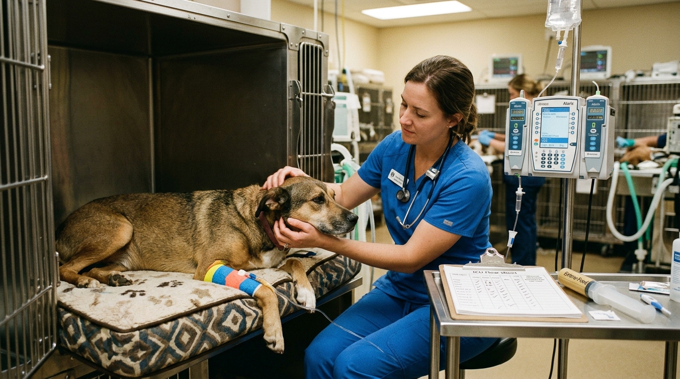
Clinical Quick Answer & Immediate Stabilization Protocol
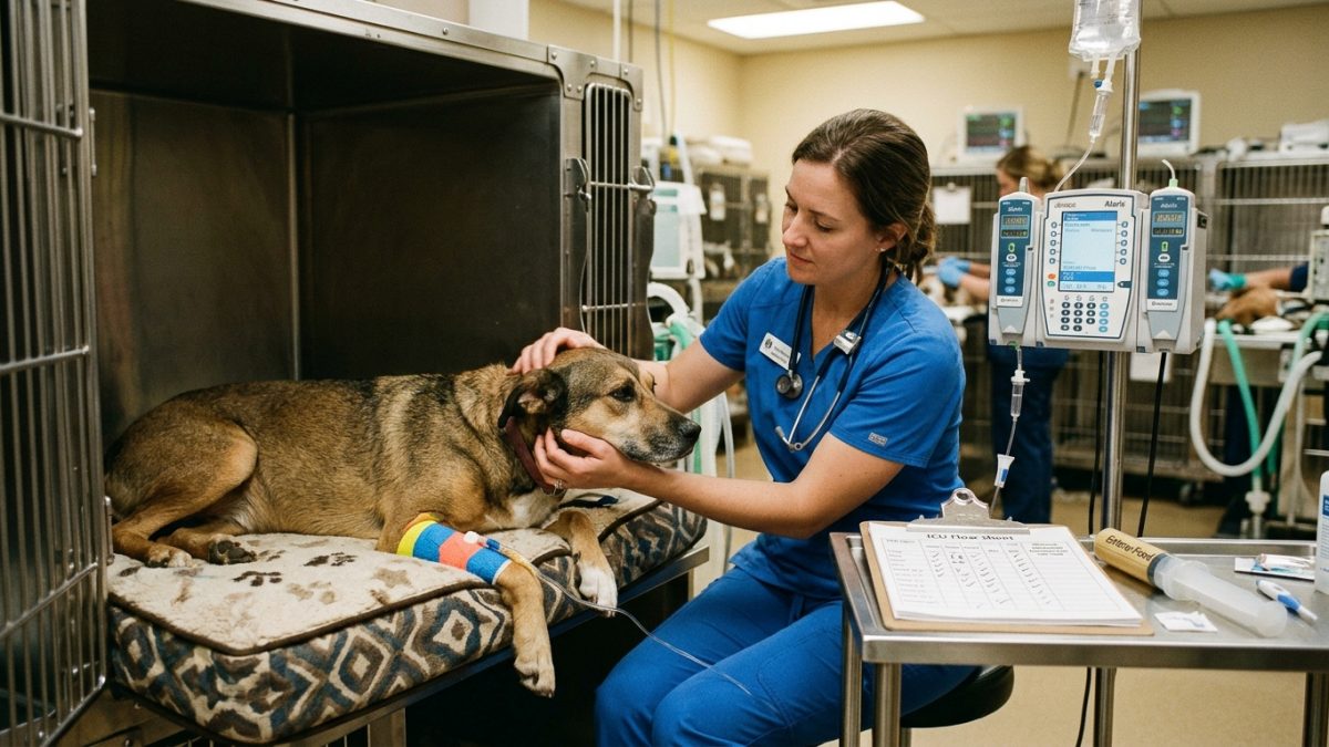
Primary Clinical Directive for Canine Diabetic Ketoacidosis (DKA) & Hyperglycemic Hyperosmolar Syndrome: A dog with polyuria and polydipsia that transitions abruptly into anorexia, vomiting, Kussmaul deep slow breathing, acetone (“nail-polish remover”) breath odor, and profound weakness has progressed from uncomplicated diabetes mellitus to Diabetic Ketoacidosis. In DKA, starving peripheral tissues force hormone-sensitive lipase to flood the liver with free fatty acids, which are oxidized into strongly acidic ketone bodies (acetoacetate, beta-hydroxybutyrate, and acetone). Hospital stabilization follows a strict sequence: (1) restore intravascular perfusion with 0.9% NaCl or balanced buffered crystalloids for 2 to 6 hours prior to insulin, (2) aggressively supplement intravenous potassium chloride and potassium phosphate, and (3) administer a low-dose continuous rate infusion (CRI) of short-acting regular crystalline insulin while screening for underlying pancreatitis or infection.
Hepatic Beta-Oxidation Ketogenesis, Osmotic Diuresis & Transcellular Potassium/Phosphorus Depletion
When insulin signaling fails and counter-regulatory glucagon dominates, hepatic glycogenolysis and gluconeogenesis drive blood glucose above the renal tubular reabsorption threshold (approx. 180–220 mg/dL in dogs). Massive glucosuria induces a relentless osmotic diuresis that flushes water, sodium, potassium, magnesium, and phosphorus into the urine. Simultaneously, unchecked hormone-sensitive lipase breaks down adipose triglycerides into non-esterified free fatty acids (FFAs). Inside hepatocyte mitochondria, elevated glucagon-to-insulin ratios activate carnitine palmitoyltransferase-1 (CPT-1), shunting FFAs exclusively into beta-oxidation and generating acetyl-CoA faster than the tricarboxylic acid (Krebs) cycle can consume it.
Excess hepatic acetyl-CoA condenses into three ketone bodies: acetoacetate, beta-hydroxybutyrate (BHB, the predominant circulating ketone in canine DKA), and acetone. Because acetoacetic and beta-hydroxybutyric acids dissociate completely at physiological blood pH, they release hydrogen ions that titrate and exhaust serum bicarbonate buffers (<15 mmol/L), producing a severe high-anion-gap metabolic acidosis. As extracellular hydrogen ions enter cells in exchange for intracellular potassium ions, serum potassium on initial admission bloodwork may appear deceptively normal despite catastrophic total-body potassium and phosphorus depletion.
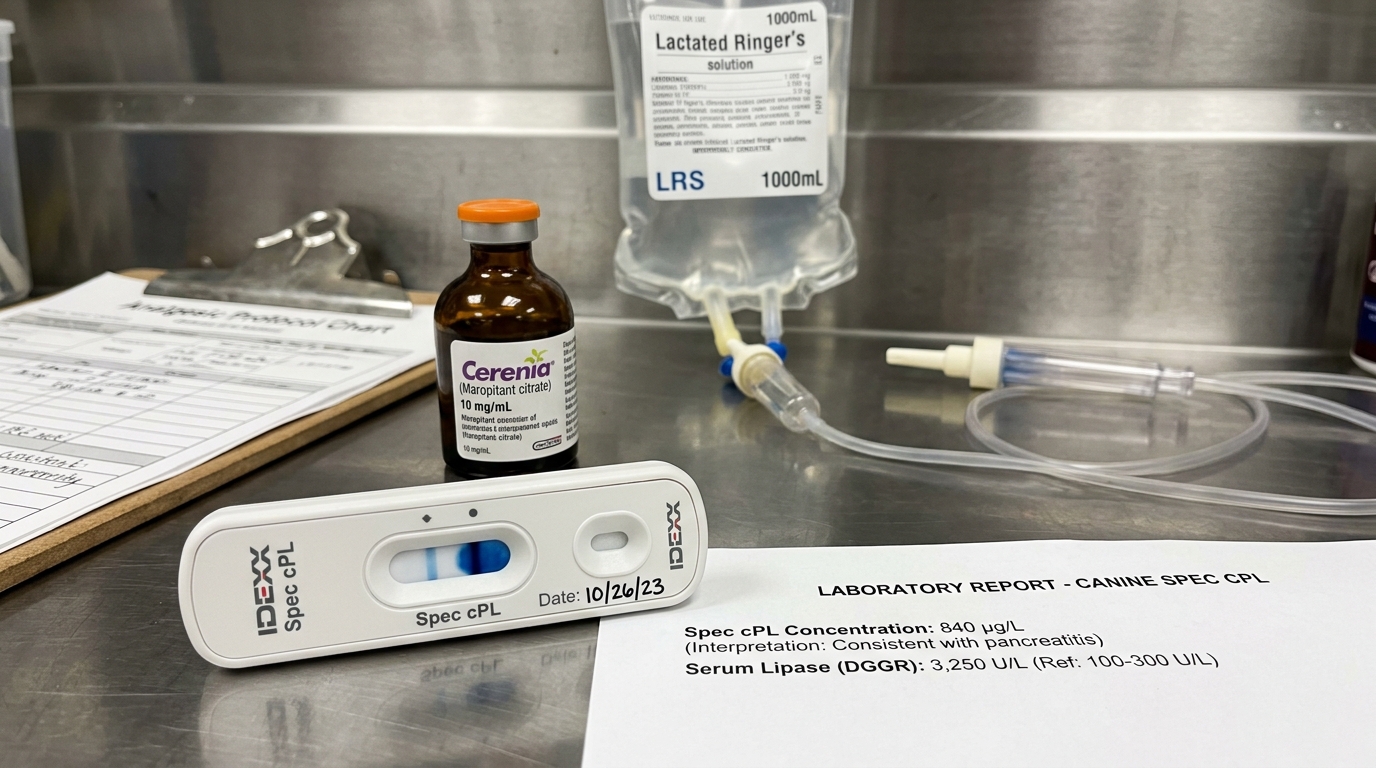
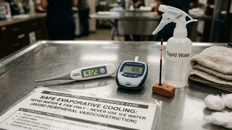
- Beta-Hydroxybutyrate (BHB) vs. Nitroprusside Urine Strips: Standard nitroprusside urine dipsticks (Ketostix) react only with acetoacetate and acetone, completely missing beta-hydroxybutyrate (the primary acid in hypoxic DKA); hand-held capillary blood BHB meters (>3.5–3.8 mmol/L) provide superior diagnostic accuracy.
- The Insulin-Driven Hypokalemic Crash: The moment intravenous regular insulin is started and acidosis begins correcting, potassium and phosphorus are driven rapidly back inside cells; without preemptive IV potassium supplementation based on sliding-scale charts, serum potassium plummets below 2.5 mmol/L, causing respiratory muscle paralysis and cardiac arrest.
- Hypophosphatemic Hemolytic Anemia: When transcellular shifts drop serum phosphorus below 1.0–1.5 mg/dL, erythrocyte 2,3-DPG and ATP levels collapse, causing acute intravascular hemolytic anemia 24 to 48 hours into DKA treatment.
- Cerebral Edema from Rapid Osmolality Drop: Lowering blood glucose faster than 50 to 75 mg/dL per hour during early resuscitation creates an osmotic gradient across the blood-brain barrier (where idiogenic osmoles persist inside neurons), drawing free water into brain parenchyma and causing fatal cerebral edema.
Emergency Triage Matrix: Clinical Presentation & Required Response
| Triage Level | Clinical Signs & Presentation | Immediate Action Protocol |
|---|---|---|
| Tier 1: Immediate Emergency (0–1 Hour) | Diabetic or polyuric dog with vomiting, refusal of food and water, sweet/fruity acetone breath, deep labored breathing (Kussmaul respiration), severe dehydration (sunken eyes, dry tacky gums), blood BHB >3.5 mmol/L, or collapse. | Withhold home insulin and transport immediately to a 24-hour veterinary ICU for central venous electrolyte monitoring, staged crystalloid volume expansion, potassium/phosphate supplementation, and regular insulin CRI. |
| Tier 2: Urgent Same-Day Evaluation (2–6 Hours) | Trace-to-moderate urine ketones or blood BHB between 1.5 and 3.0 mmol/L in a diabetic dog that is still bright, alert, hydrated, and willing to eat without vomiting (“Diabetic Ketosis”). | Contact your veterinarian immediately for a same-day outpatient workup (urinalysis with culture, Spec cPL pancreatitis test, electrolyte panel) to adjust insulin and hydration before acidosis develops. |
| Tier 3: Post-DKA Transition to Subcutaneous Insulin | Hospitalized DKA survivor that is hydrated, non-vomiting, eating voluntarily for >24 hours, with resolved ketonemia (BHB <0.6 mmol/L) and normalized venous pH/bicarbonate. | Transition back to twice-daily subcutaneous intermediate-acting insulin (Vetsulin/NPH) with consistent high-fiber/low-fat meals, overlapping the final IV regular insulin hours under veterinary supervision. |
Staged Fluid Resuscitation, Sliding-Scale Potassium/Phosphate & Comorbidity Eradication
A foundational rule of veterinary critical care is that intravenous regular insulin is delayed for the first 2 to 6 hours of DKA hospitalization while isotonic crystalloid fluids (0.9% NaCl, Normosol-R, or Plasmalyte-148) restore glomerular filtration and tissue perfusion. Fluid expansion alone reduces blood glucose by 30 to 50 percent through hemodilution and renal urinary glucose clearance while suppressing counter-regulatory epinephrine and cortisol levels. Starting insulin in a hypovolemic dog before intravascular volume and baseline serum potassium are restored shifts water and potassium intracellularly, precipitating circulatory shock and lethal arrhythmia.
Once initial rehydration is underway and serum potassium is confirmed above 3.5 mmol/L, low-dose short-acting regular crystalline insulin (0.05 to 0.09 U/kg/day via continuous intravenous syringe pump or hourly intramuscular protocol) is initiated to halt hepatic ketogenesis. As blood glucose declines below 250 mg/dL, 2.5% to 5.0% dextrose is added to the intravenous fluids so that the regular insulin infusion can continue uninterrupted until all circulating ketone bodies and metabolic acidosis are completely cleared—a process that typically takes 36 to 72 hours longer than lowering blood glucose.
- Mandatory Search for the Triggering Comorbidity: Every DKA workup requires a sterile cystocentesis urine culture (as glucosuria and immunocompromise mask subclinical pyelonephritis), canine pancreatic lipase immunoreactivity (Spec cPL), abdominal ultrasound, and thoracic radiographs.
- Ovariohysterectomy in Intact Female Diabetics: In unspayed female dogs, luteal-phase progesterone stimulates mammary growth hormone secretion, causing severe insulin resistance and DKA; emergency ovariohysterectomy is mandatory once the patient is metabolically stabilized.
- Sliding-Scale Potassium & Phosphate Protocol: Serum electrolytes (K+, PO4, Na+, Cl-) are rechecked every 4 to 6 hours; when serum phosphorus drops below 2.0 mg/dL, half of the potassium requirement is delivered as potassium phosphate (0.03–0.06 mmol/kg/hr) alongside potassium chloride.
- Never Stop Insulin Simply Because Glucose Normalizes: In DKA, insulin is administered to shut off hormone-sensitive lipase and ketogenesis, not merely to lower glucose; when blood sugar reaches 200–250 mg/dL, clinicians add IV dextrose rather than turning off the insulin pump until ketonemia resolves.
Strict Rule S1 Clinical Red Flags: Canine Diabetic Ketoacidosis (DKA) & Hyperglycemic Hyperosmolar Syndrome
Critical safety boundaries for Canine (Canis lupus familiaris — Newly Diagnosed & Decompensated Diabetic Dogs) caregivers:
- Zero Full-Dose Home Insulin When a Diabetic Dog Refuses Food: Never inject your dog’s full prescribed maintenance dose of Vetsulin or NPH insulin if the dog refuses its meal or is actively vomiting; contact your veterinarian immediately for sick-day triage and ketone testing.
- Zero Rapid Subcutaneous Boluses of Long-Acting Insulin for Ketones: Never attempt to clear positive urine ketone strips at home by injecting extra units of long-acting insulin, as depot accumulation causes delayed severe hypoglycemia without resolving electrolyte depletion.
Clinical Preparation & Biosecurity Action Checklist
- Keep a pack of fresh, foil-sealed urine ketone test strips (or a capillary blood beta-hydroxybutyrate ketone meter) in your home diabetic care kit.
- Test for urine or blood ketones immediately any time your diabetic dog skips a meal, vomits, seems lethargic, or drinks excessively for more than 24 hours.
- Bring your home insulin vial (checking the expiration date, storage temperature, and gentle rolling vs. shaking technique) and syringe size (U-40 vs. U-100) to the emergency hospital.
- Feed a consistent, weighed ultra-low-fat, high-complex-carbohydrate diet at the exact same 12-hour intervals following post-DKA hospital discharge.
Related Clinical Guides & Triage Protocols
Peer-Reviewed Clinical References & Evidence Grounding
- 2018 AAHA Diabetes Management Guidelines for Dogs and Cats (opens in new tab) (Journal of the American Animal Hospital Association (2018) — AAHA Consensus Guidelines by Behrend et al. indexed on NCBI PubMed (PMID: 29446226).)
- Evaluation of a continuous rate infusion of regular insulin for treatment of diabetic ketoacidosis in dogs (opens in new tab) (Journal of Veterinary Emergency and Critical Care (2012) — Peer-reviewed clinical trial indexed on NCBI PubMed (PMID: 22748169).)
- Diagnostic utility of a point-of-care beta-hydroxybutyrate sensor for diabetic ketosis and ketoacidosis in dogs and cats (opens in new tab) (Journal of Veterinary Internal Medicine (2008) — Peer-reviewed diagnostic accuracy study indexed on NCBI PubMed (PMID: 18489578).)
- Merck Veterinary Manual: Diabetes Mellitus and Diabetic Ketoacidosis in Dogs and Cats (opens in new tab) (Verified veterinary clinical reference on canine DKA fluid resuscitation, potassium/phosphorus repletion, and insulin CRI protocols.)
Source transparency
Reference mix for this guide
Source labels classify the institutional category of the references cited above.