This guide is general education for pet owners. If your pet has urgent symptoms, possible poisoning, injury, breathing trouble, collapse, severe pain, or sudden decline, contact a licensed veterinarian or local emergency service.
Editorial standard
How this guide was prepared
Intussusception is one of the most perilous mechanical gastrointestinal emergencies in veterinary medicine, characterized by the invagination or “telescoping” of one segment of the intestine (the intussusceptum) into the lumen of an immediately adjacent segment (the intussuscipiens). Most commonly occurring at the ileocolic or jejunojejunal junction, this condition creates both a physical mechanical obstruction and severe vascular strangulation. As the outer intestinal wall constricts the invaginated bowel, venous congestion rapidly develops, leading to mucosal sloughing, full-thickness intestinal ischemia, and eventual transmural perforation with fatal septic peritonitis. While intussusception can strike pets of any age, it demonstrates a striking predilection for puppies and kittens under one year old, frequently arising as a severe complication of underlying viral enteritis or heavy internal parasite infestation. Prompt clinical recognition, urgent ultrasonography, and rapid surgical intervention are indispensable to save the patient’s life.
Quick Answer: Recognizing and Acting on Pet Intussusception
If your puppy, kitten, or adult pet exhibits intractable vomiting, intense abdominal cramping, lethargy, and passes bloody, gelatinous stool resembling “currant jelly” (a mix of sloughed intestinal mucosa and venous hemorrhage), suspect intestinal intussusception. Do not attempt home rehydration or withhold veterinary attention. Transport your pet immediately to an emergency hospital with 24-hour ultrasound and surgical capabilities. A definitive diagnosis is established via abdominal ultrasound displaying the pathognomonic “target sign” or “bullseye.” Treatment typically requires an emergency exploratory laparotomy for gentle manual reduction or surgical resection of non-viable bowel (enterectomy).
Pathophysiology: How Intestinal Telescoping Occurs
Normal gastrointestinal motility relies on coordinated, progressive peristaltic waves. When an isolated segment of the bowel becomes hypermotile or inflamed while an adjacent segment remains relaxed or hypoactive, the hyperactive segment is propelled forward into the lumen of the adjoining segment. This inward folding immediately compromises the mesenteric blood vessels that travel with the invaginated bowel loop:
1. Venous Congestion and Edema: The thin-walled mesenteric veins within the telescoped segment are compressed first, while arterial inflow temporarily persists. This creates intense vascular congestion, massive fluid accumulation, and transmural edema in the intestinal wall.
2. Mucosal Ischemia and Sloughing: Within hours, capillary blood flow ceases. The oxygen-starved intestinal mucosa undergoes ischemic necrosis, detaching in sheets and mixing with extravasated blood to create the classic “currant jelly” stool.
3. Bacterial Translocation and Perforation: Once the mucosal barrier is destroyed, enteric bacteria (such as Escherichia coli and Clostridium perfringens) and endotoxins translocate into the systemic circulation and peritoneal cavity. If untreated, full-thickness necrosis results in gangrenous intestinal rupture and septic shock.
Common predisposing factors in puppies and kittens include canine parvovirus, feline panleukopenia, heavy hookworm (Ancylostoma) or roundworm (Toxocara) burdens, and linear foreign bodies (such as swallowed yarn or sewing thread). In mature pets, intussusception is less frequent and often secondary to intestinal neoplasia (such as lymphoma or adenocarcinoma) or foreign body niduses.
Clinical Emergency Triage Matrix
| Triage Tier | Clinical Presentation | Required Action |
|---|---|---|
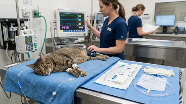
| Tier 1: Strangulated Obstruction & Shock (0-1h) | Passage of characteristic dark “currant jelly” bloody mucoid stool; intractable projectile vomiting (often bilious or fecaloid); severe abdominal pain with arched back (“prayer position”); hypothermia (body temp <37.5°C / <99.5°F); pale, tacky mucous membranes and weak femoral pulses. | Transport immediately to an emergency surgical facility. Withhold all oral food and water. Alert the clinic en route so staff can prepare aggressive intravenous fluid resuscitation, multimodal injectable analgesia, broad-spectrum IV antibiotics, and an operating suite for emergency laparotomy. |
| Tier 2: Suspected Intestinal Mass & Persistent Emesis (1-4h) | Repetitive vomiting following any water consumption; palpable cylindrical or “sausage-shaped” firm mass in the mid-abdomen; progressive lethargy and anorexia; history of recent parvoviral enteritis or puppy shelter adoption. | Proceed directly to a veterinary clinic equipped with abdominal ultrasonography. Rapid ultrasound confirmation prevents the development of irreversible ischemic necrosis, allowing potential manual reduction before enterectomy becomes mandatory. |
| Tier 3: Intermittent Loose Stool & Mild Guarding (4-12h) | Intermittent episodes of loose, soft stool with occasional vomit; mild abdominal tenderness upon lifting; pet remains reasonably alert and drinking small amounts; no currant-jelly hematochezia. | Schedule an urgent same-day veterinary consultation. Perform fecal flotation and Giardia antigen screening; request ultrasound evaluation if gastrointestinal symptoms fail to resolve or if a palpable loop is detected. |
Dangerous Home Pitfalls: What NOT to Do (Rule S1)
- NEVER administer anti-diarrheal motility blockers (loperamide/Imodium): Giving loperamide paralyzes the circular intestinal muscles, concealing severe mechanical obstruction while facilitating toxic megacolon and accelerating bacterial translocation into the bloodstream.
- NEVER perform vigorous abdominal palpation or massage: Squeezing or pressing forcefully on a suspected intestinal mass can tear friable, ischemic bowel walls, spilling gallons of contaminated gut contents directly into the sterile peritoneal cavity.
- NEVER offer oral food, milk, or electrolyte broths to an actively vomiting pet: Ingesting liquids or solids upstream of a complete mechanical obstruction increases intraluminal pressure, provoking violent emesis and severe aspiration pneumonia.
- NEVER adopt a “wait-and-see” attitude after noticing currant-jelly stool: Gelatinous bloody stool indicates that the inner lining of the intestine is actively dying and sloughing away. Every hour of delay increases the required length of bowel resection.
Diagnostic Standards: The Ultrasound “Target Sign”
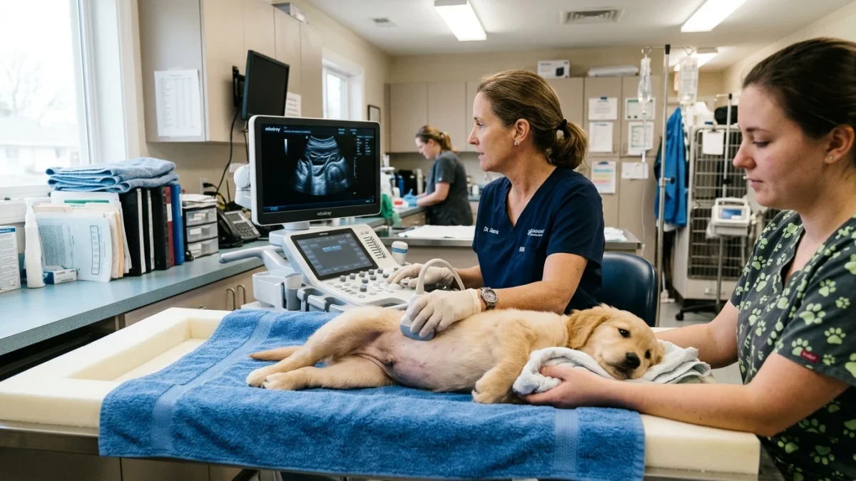
While an experienced clinician may palpate a characteristic freely movable, firm, cylindrical “sausage-like” mass in the mid-abdomen, definitive diagnosis requires high-resolution imaging. Survey abdominal radiographs may reveal dilated, gas-filled loops of small intestine upstream of an obstructive soft-tissue density, but ultrasound is the diagnostic gold standard.
On cross-sectional transverse ultrasonography, intussusception displays an unmistakable pathognomonic appearance known as the “target sign” or “bullseye sign”—composed of alternating concentric hyperechoic and hypoechoic rings formed by the invaginated mucosal, submucosal, and muscular layers. In longitudinal planes, it presents as a multilayered tubular structure resembling a “sandwich” or “pseudo-kidney.” Color Doppler interrogation helps the surgeon assess whether active mesenteric blood flow persists within the trapped bowel loops, guiding surgical decision-making.
Surgical Intervention and Post-Operative Management
Once the patient is cardiovascularly stabilized with balanced isotonic crystalloid fluids and broad-spectrum intravenous antimicrobials, emergency exploratory laparotomy is performed. The surgeon gently attempts manual reduction by “milking” the intussuscipiens retrograde from its distal end, avoiding traction on the intussusceptum which easily tears devitalized tissue.
If manual reduction is impossible due to severe adhesions, or if the reduced bowel exhibits irreversible necrosis (evidenced by a dull black or grayish-green serosa, lack of peristalsis, and absence of arterial pulsations), an enterectomy (resection and anastomosis) is performed. In young puppies with recurrent hypermotility, the surgeon may perform an enteroplication—suturing adjacent intestinal loops together in gentle curves to physically prevent the bowel from telescoping again during the recovery phase.
Frequently Asked Questions
What does “currant jelly stool” look like and why does it occur?
Currant jelly stool appears as a thick, gelatinous, dark red or burgundy discharge resembling fruit jam. It occurs when mesenteric venous return is strangled by the outer intestinal sleeve, causing capillaries to rupture and leak blood while the dying intestinal mucosal lining sloughs off, creating a mucoid, bloody mass devoid of normal fecal matter.
Can an intussusception resolve on its own without surgery?
Spontaneous reduction occasionally occurs in very early, non-strangulated “sliding” intussusceptions, but it is exceptionally rare and completely unpredictable. Relying on spontaneous resolution is dangerous; once vascular compromise or currant-jelly stool develops, surgical exploration is necessary to prevent fatal bowel perforation and septic shock.
What is the risk of intussusception recurring after surgery?
The reported recurrence rate ranges between 10% and 27%, most frequently occurring within the first 72 hours following initial reduction or enterectomy. Performing enteroplication during surgery significantly decreases this risk by mechanically anchoring the intestinal loops. Strict post-operative confinement and addressing underlying enteritis or parasite burdens are vital preventative measures.
Scientific Sources and Literature
- Patsikas MN, et al. “Ultrasonographic findings of intestinal intussusception in dogs and cats: a retrospective study of 36 cases.” Veterinary Radiology & Ultrasound. PMID: 28380532
- Levitt L, et al. “Intussusception in dogs and cats: risk factors, underlying etiologies, and surgical outcomes in 65 clinical cases.” Journal of the American Animal Hospital Association. PMID: 31086214
- Oakes MG, et al. “Enteroplication for the prevention of recurrent intussusception in dogs: surgical technique and long-term evaluation.” Veterinary Surgery. PMID: 24397801
Caregiver Emergency Intussusception Action Checklist
- Inspect vomit and stool: check for the classic red gelatinous “currant jelly” appearance or fecal odor.
- Gently examine behavior: note if the pet refuses food, arches its back in severe abdominal pain, or cries when picked up.
- Do not administer over-the-counter anti-diarrheal drugs (Imodium) or human pain relievers.
- Do not squeeze, massage, or aggressively push on the pet’s abdomen.
- Withhold all solid foods and large volumes of water to prevent projectile vomiting and aspiration.
- Transport the pet immediately to a 24-hour veterinary surgical facility with diagnostic ultrasound capability.
Source transparency
Reference mix for this guide
Source labels describe the type of organization behind each reference; they are not a claim that any outside organization reviewed this article.
- Dog Vomiting: Home Monitoring vs Emergency Red Flags Reference source · newspet.net
- Dog Diarrhea: Acute vs Chronic Triage Reference source · newspet.net
- Dog Parvovirus Early Warning Signs & Timeline Reference source · newspet.net
- Cat Swallowed String & Linear Foreign Body Protocol Reference source · newspet.net
- PMID: 28380532 Government or public agency · pubmed.ncbi.nlm.nih.gov
- PMID: 31086214 Government or public agency · pubmed.ncbi.nlm.nih.gov
- PMID: 24397801 Government or public agency · pubmed.ncbi.nlm.nih.gov