This guide is general education for pet owners. If your pet has urgent symptoms, possible poisoning, injury, breathing trouble, collapse, severe pain, or sudden decline, contact a licensed veterinarian or local emergency service.
Editorial standard
How this guide was prepared
Sudden collapse accompanied by chalky white or pale mucous membranes in a dog is one of the most urgent, critical presentations encountered in veterinary emergency medicine. This dramatic clinical presentation frequently signals catastrophic acute internal hemorrhage into the peritoneal cavity, known clinically as hemoperitoneum. In middle-aged to geriatric large-breed dogs, spontaneous internal bleeding is most often triggered by the sudden rupture of a vascular splenic mass—either a benign splenic hematoma or a malignant hemangiosarcoma. When the splenic capsule or vascular tumor ruptures, substantial arterial blood pours directly into the abdominal cavity, precipitating severe hypovolemic shock, critical tissue hypoperfusion, and cellular hypoxia within minutes.
Quick Answer: First Aid for Sudden Canine Collapse and Pale Gums
If your dog suddenly collapses, becomes weak, or cannot stand, immediately lift the upper lip to inspect the gums. If the gums are porcelain white, light pink, or gray instead of healthy salmon pink, your dog is in acute circulatory shock—most commonly from internal abdominal hemorrhage (ruptured spleen) or cardiac tamponade. Keep your dog warm and flat; avoid lifting by the stomach. Transport your dog immediately to the nearest 24-hour veterinary emergency center for focused abdominal ultrasound (AFAST), intravenous blood volume resuscitation, and emergency surgical stabilization.
Evaluating Mucous Membrane Color and Femoral Pulse Deficits
Recognizing the physical signs of hypovolemic shock allows caregivers to distinguish acute systemic circulatory collapse from simple musculoskeletal slips or localized limb pain. The primary diagnostic window is the oral mucous membrane. In a healthy, well-oxygenated dog, the gums above the canine teeth display a rich salmon-pink color, and when pressed with a finger, the blanched spot refills with pink blood within 1 to 2 seconds (capillary refill time, or CRT). In dogs experiencing active intra-abdominal hemorrhage, the gums turn ghostly pale, porcelain white, or grayish-blue. The capillary refill time is either severely prolonged (>3 seconds) or completely sluggish, indicating massive peripheral vasoconstriction as the body attempts to shunt remaining blood to the heart and brain.
Palpating the femoral pulse along the medial thigh provides another critical clinical clue. As intravascular volume plummets from internal hemorrhage, the femoral arterial pulse becomes exceedingly rapid (tachycardia >140 to 180 beats per minute) and feels weak, thready, or difficult to detect. The dog’s extremities—including paws, ear flaps, and muzzle—turn distinctly cold to the touch as peripheral blood vessels constrict.
Clinical Emergency Triage Matrix
| Triage Tier | Clinical Presentation | Required Action |
|---|---|---|
| Tier 1: Immediate Critical Emergency (0-1h) | Sudden recumbency and inability to rise; chalky white or muddy gums; distended, fluid-filled abdomen with visible fluid wave; weak or undetectable femoral pulse; rapid shallow panting or agonal respirations. | Call the emergency hospital immediately to notify them of incoming hemorrhagic shock. Transport dog on a flat blanket or board stretcher without abdominal compression. Prepare for immediate emergency venous access and blood transfusion. |
| Tier 2: Urgent Veterinary Triage (1-4h) | Episodic weakness where the dog stumbled or collapsed briefly, appeared dazed, but subsequently stood back up; gums pale pink; mild abdominal tenderness or reluctance to lie down; heart rate moderately elevated. | Proceed directly to an emergency veterinary hospital. Transient recovery occurs when blood pressure drops low enough for temporary clot formation; re-bleeding frequently follows within hours without surgical hemostasis. |
| Tier 3: Same-Day Evaluation (4-8h) | Mild lethargy and slow movement without collapse; gums remain pink with normal CRT (<2 seconds); strong femoral pulses; no abdominal distension or respiratory distress. | Restrict physical exertion completely. Schedule an urgent outpatient veterinary visit for complete blood counts, hematocrit/packed cell volume (PCV) evaluation, and abdominal palpation. |
Dangerous Home Pitfalls: What NOT to Do (Rule S1)
- NEVER squeeze or lift the dog around the abdomen: When transporting a collapsed large dog, caregivers frequently attempt to lift them by scooping under the belly. Placing mechanical pressure on an enlarged, ruptured spleen ruptures protective hematoma seals, causing an immediate, fatal surge of active internal bleeding. Always lift by supporting the bony chest and pelvis.
- NEVER apply tight abdominal wraps or compression bands: Compressive home wrapping restricts diaphragmatic excursion, triggering severe hypoventilation and respiratory arrest while failing to halt high-pressure arterial splenic bleeding.
- NEVER administer human or pet pain medications: Non-steroidal anti-inflammatory drugs (NSAIDs) and aspirin inhibit cyclooxygenase enzymes and interfere with platelet aggregation. In the setting of active internal hemorrhage, giving these medications destroys the body’s natural coagulation cascade.
- NEVER ignore a “temporary recovery” after an episodic collapse: When internal hemorrhage occurs, systemic blood pressure drops precipitously, allowing a temporary clot to form over the ruptured vessel and permitting the dog to temporarily stand back up. This “honeymoon period” is deceptive; as blood pressure normalizes, the clot inevitably dislodges, triggering fatal secondary collapse.
Pathophysiology: Splenic Masses, Hemangiosarcoma, and Trauma
The canine spleen is an extremely vascular organ located in the left cranial abdomen, functioning as a primary reservoir for red blood cells and a filtration organ for the immune system. Splenic masses occur with high frequency in older dogs, particularly breeds like German Shepherds, Golden Retrievers, Labrador Retrievers, and Boxers. Veterinary clinical data demonstrates the classic “two-thirds rule”: approximately two-thirds of all canine splenic masses are malignant, and of those malignant tumors, roughly two-thirds are hemangiosarcomas—an aggressive, highly invasive cancer originating from endothelial cells lining blood vessels.
Even benign splenic masses, such as massive splenic hematomas or nodular lymphoid hyperplasia, pose an imminent life hazard. Because the splenic tissue becomes expanded and structurally fragile, mild physical activity—such as jumping out of a car, playing fetch, or rolling in the yard—can shear the exterior capsule. When this occurs, hundreds of milliliters of blood rapidly pool into the peritoneal space. As circulating volume drops, oxygen delivery to coronary and cerebral vessels fails, culminating in hypovolemic cardiovascular shock.
Frequently Asked Questions
How does an emergency veterinarian diagnose internal bleeding in a collapsed dog?
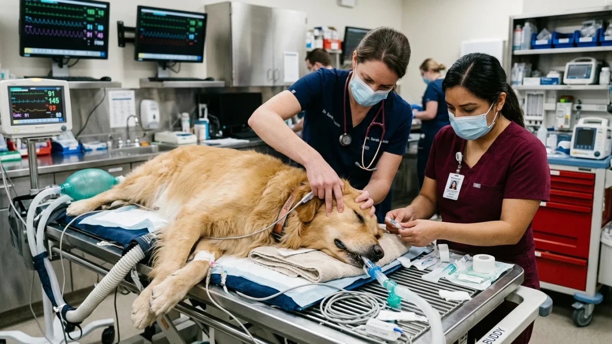
An emergency clinician immediately performs an abdominal focused assessment with sonography for trauma (AFAST ultrasound). This rapid point-of-care scan takes less than 60 seconds to visualize pockets of free fluid around the liver, spleen, and bladder. If fluid is identified, the clinician performs an abdominocentesis—inserting a sterile needle into the abdomen to aspirate fluid. If non-clotting, whole blood is retrieved, a definitive diagnosis of active hemoperitoneum is confirmed.
What emergency treatment is required to save a dog with a ruptured spleen?
Treatment begins with rapid intravenous resuscitation using isotonic crystalloids and synthetic colloids via large-bore IV catheters, often accompanied by crossmatched whole blood or packed red blood cell transfusions to stabilize blood pressure. Once cardiovascular stability is achieved, the dog undergoes emergency exploratory laparotomy and emergency splenectomy—surgical removal of the entire spleen and ligation of splenic blood vessels to permanently halt the internal hemorrhage.
Can a dog live a normal life after having their spleen surgically removed?
Yes, dogs can live a full, normal life without a spleen, as the liver and bone marrow assume its functions of filtering blood and recycling aged red blood cells. The long-term prognosis depends entirely on the histological biopsy of the removed splenic mass. If the mass is a benign hematoma, emergency surgical splenectomy is 100 percent curative. If the mass is confirmed as malignant hemangiosarcoma, adjuvant chemotherapy and oncological management are recommended to extend quality of life.
Scientific Sources and Literature
- Pintar J, et al. “Acute hemoperitoneum in dogs: a retrospective review of 67 cases (1999-2001).” Journal of the American Animal Hospital Association. PMID: 26362842
- Boysen SR, Lisciandro GR. “The use of ultrasound for dogs and cats in the emergency room: AFAST and TFAST.” Veterinary Clinics of North America: Small Animal Practice. PMID: 28987115
- Lynch AM, et al. “Transfusion practices for treatment of dogs with hemoperitoneum: 73 cases (2007-2012).” Journal of Veterinary Emergency and Critical Care. PMID: 30129188
Caregiver Emergency Action Checklist for Canine Collapse
- Inspect oral mucous membranes immediately: check whether gums are healthy pink or dangerously white, gray, or pale.
- Measure capillary refill time: press firmly on the gum line above the canine tooth and count seconds until color returns.
- Assess extremities: feel paws and ears to check for abnormal coldness and lack of peripheral warmth.
- Avoid applying pressure or lifting under the abdomen; use a blanket or board stretcher supported under the chest and rump.
- Keep the dog horizontally flat and gently cover with a warm blanket to conserve body temperature during transit.
- Call the emergency veterinary facility en route so the trauma team can prepare oxygen, ultrasound, and blood products prior to arrival.
Source transparency
Reference mix for this guide
Source labels describe the type of organization behind each reference; they are not a claim that any outside organization reviewed this article.
- Dog Bleeding Wound & Hemorrhage First Aid Reference source · newspet.net
- Dog Shaking & Lethargic Differential Triage Reference source · newspet.net
- Dog Bloat & GDV Gastric Torsion Emergency Reference source · newspet.net
- Dog Hypothermia & Active Rewarming Triage Reference source · newspet.net
- PMID: 26362842 Government or public agency · pubmed.ncbi.nlm.nih.gov
- PMID: 28987115 Government or public agency · pubmed.ncbi.nlm.nih.gov
- PMID: 30129188 Government or public agency · pubmed.ncbi.nlm.nih.gov