This guide is general education for pet owners. If your pet has urgent symptoms, possible poisoning, injury, breathing trouble, collapse, severe pain, or sudden decline, contact a licensed veterinarian or local emergency service.
Editorial standard
How this guide was prepared
Gastric Dilatation-Volvulus (GDV), commonly known as dog bloat, is the single most time-critical surgical emergency in veterinary medicine. Without immediate surgical intervention, the mortality rate approaches 100 percent. The condition involves the rapid accumulation of gas within the stomach followed by physical rotation along its axis, obstructing blood flow and leading to systemic shock, tissue necrosis, and cardiovascular collapse within hours. Understanding the mechanics of this catastrophic event is essential for providing the immediate response necessary to maximize survival probability.
Quick Answer: Is Dog Bloat an Emergency?
Yes. Dog bloat (GDV) is a catastrophic medical emergency requiring immediate surgical intervention. The hallmark signs include repeated, non-productive retching or gagging (attempting to vomit without producing anything), sudden expansion of the abdomen which feels hard to the touch, and extreme restlessness with an inability to settle. If you observe these clinical signs, immediate transport to an emergency veterinary hospital is required. Do not wait to see if the symptoms resolve. Time to surgery is the primary determinant of survival.
Pathophysiology: Understanding Gastric Dilatation-Volvulus
The term GDV describes a two-part pathological process. Gastric dilatation refers to the abnormal accumulation of gas, fluid, or food within the stomach, causing it to expand far beyond its normal physiological capacity. Volvulus occurs when this distended stomach rotates or twists on its mesenteric axis. This torsion seals off both the entry (cardia) and exit (pylorus) of the stomach, trapping the contents and continuing to increase intragastric pressure at a terrifying rate. The exact initiating trigger for this process remains an area of ongoing veterinary research, though aerophagia (swallowing excess air) and delayed gastric emptying are prominent physiological precursors.
As the stomach expands, it exerts mechanical pressure on the caudal vena cava, the large vein returning deoxygenated blood to the heart. This severely restricts venous return, precipitating a massive reduction in cardiac output and triggering hypovolemic shock. Simultaneously, the twisting of the stomach compromises its own blood supply, initiating rapid tissue ischemia and necrosis (death of the stomach wall). The compromised blood flow also affects the spleen, which is anatomically linked and often rotates alongside the stomach. This rotation leads to splenic congestion, ischemia, and the release of myocardial depressant factors and endotoxins into the bloodstream, triggering systemic inflammatory response syndrome (SIRS) and disseminated intravascular coagulation (DIC). The entire physiological cascade occurs rapidly, often transitioning from initial symptoms to irreversible cardiovascular collapse within a two-hour window.
Dangerous Home Pitfalls: What NOT to Do (Rule S1)
- NEVER attempt to relieve gastric gas at home by passing tubes down the esophagus; this can cause esophageal rupture or delay transport.
- NEVER administer over-the-counter anti-gas medications (such as simethicone or Gas-X) as a primary treatment; these do not resolve a volvulus and provide a false sense of security while critical tissue dies.
- NEVER adopt a “wait and see” approach when a deep-chested dog exhibits non-productive retching. Time lost is tissue lost.
- NEVER allow a dog to engage in vigorous exercise, running, or high-intensity play within one hour of consuming a large volume of food or water.
Clinical Triage Matrix: Evaluating Abdominal Symptoms
| Triage Tier | Clinical Presentation | Required Action |
|---|---|---|
| Tier 1: Immediate Emergency (0-2h) | Non-productive retching/gagging with nothing coming up; visibly distended, rock-hard abdomen; extreme restlessness, pacing, excessive drooling, inability to lie down comfortably; rapid shallow breathing with a distended belly; collapse after unproductive vomiting attempts. | Proceed to nearest veterinary emergency hospital immediately. Notify them you are en route with a suspected GDV. Do not hesitate. |
| Tier 2: Same-Day Veterinary Assessment (12-24h) | Excessive drooling accompanied by mild abdominal discomfort; pacing and whining after consuming a large meal; a single unproductive retch that subsequently resolves without further symptoms. | Schedule an immediate veterinary evaluation. Do not feed further meals until cleared by a veterinarian to ensure partial torsion or early dilatation is not occurring. |
| Tier 3: Home Monitoring | Brief post-meal belly gurgling with normal behavior; occasional burping; normal energy levels and comfortable resting position. | Monitor behavior. If symptoms escalate to discomfort or retching, upgrade to Tier 1 or Tier 2. Ensure adequate rest post-feeding. |
The Progression Timeline of GDV
The timeline of GDV is aggressive and unforgiving. Recognizing the progression stages can facilitate earlier intervention, although transit to a veterinary facility should occur immediately upon suspecting the condition regardless of the presumed stage.
0 to 30 Minutes: Initial Dilatation and Discomfort
The earliest signs involve subtle behavioral changes. The dog will typically appear anxious, pacing the room, and unable to find a comfortable resting position. Hypersalivation (excessive drooling) begins as nausea increases. The dog may look repeatedly at their flank or abdomen, indicating localized visceral pain. Abdominal distension may not be visually obvious at this stage, particularly in heavily coated or overweight breeds, but the stomach is actively expanding and tension is building.
30 to 60 Minutes: Volvulus and Shock Onset
As the stomach twists, the mechanical obstruction is complete. The hallmark symptom of continuous, non-productive retching emerges. The dog will forcefully attempt to vomit, producing only strings of thick saliva or white foam, but no actual stomach contents. The abdomen becomes visibly enlarged, taut, and tympanic (sounding like a hollow drum if tapped gently). Cardiac output plummets due to vena cava compression. The heart rate spikes (tachycardia) as the body attempts to maintain systemic blood pressure, while the mucous membranes (gums) may turn pale, gray, or muddy, indicating poor peripheral perfusion. The respiratory rate increases dramatically as the distended stomach physically impedes the diaphragm, limiting lung capacity and creating severe respiratory distress.
60 to 120 Minutes: Tissue Necrosis and Systemic Collapse
Without immediate surgical and medical intervention, ischemic necrosis of the gastric wall and spleen rapidly advances. The profound lack of oxygenated blood causes full-thickness tissue death, leading to the potential rupture of the stomach wall. Gastric rupture results in catastrophic, invariably fatal peritonitis. Severe cardiac arrhythmias, particularly ventricular premature complexes (VPCs), are extremely common during this phase due to myocardial ischemia and severe endotoxemia. The dog will typically progress from frantic restlessness to extreme lethargy, lateral recumbency (collapse), and eventually loss of consciousness. Resuscitation efforts at this advanced stage are significantly more complex, and the prognosis declines precipitously with every passing minute.
Risk Factors for GDV
While GDV can theoretically occur in any canine, specific anatomical, genetic, and environmental factors drastically increase individual susceptibility.
- Anatomy and Breed Conformation: Deep-chested breeds (those with a large thoracic depth to width ratio) are heavily predisposed. Primary at-risk breeds include the Great Dane, German Shepherd Dog, Standard Poodle, Weimaraner, Saint Bernard, Gordon Setter, Irish Setter, and Doberman Pinscher. The anatomical space allows for greater mobility of the stomach within the abdominal cavity.
- Feeding Practices: Consuming a single, very large meal daily significantly increases the mechanical weight and volumetric risk compared to feeding two or more smaller, portion-controlled meals. Rapid consumption of food (speed eating) introduces significant aerophagia (swallowing massive amounts of air).
- Elevated Feeding: Historically recommended as a preventative measure, robust clinical studies have demonstrated that using elevated food bowls actually increases the statistical risk of GDV, potentially by facilitating faster eating and increased air ingestion.
- Stress and Temperament: Dogs with highly anxious, fearful, or reactive temperaments, or those experiencing acute environmental stress (such as boarding, travel, or major routine disruptions), exhibit notably higher incidence rates of GDV.
- Age and Genetics: Risk increases consistently with age, particularly in dogs over seven years old due to increasing laxity of the hepatogastric ligaments holding the stomach in place. Furthermore, having a first-degree relative (parent or sibling) with a history of GDV significantly elevates a dog’s individual genetic risk profile.
Veterinary Diagnostics and Surgical Intervention Protocols
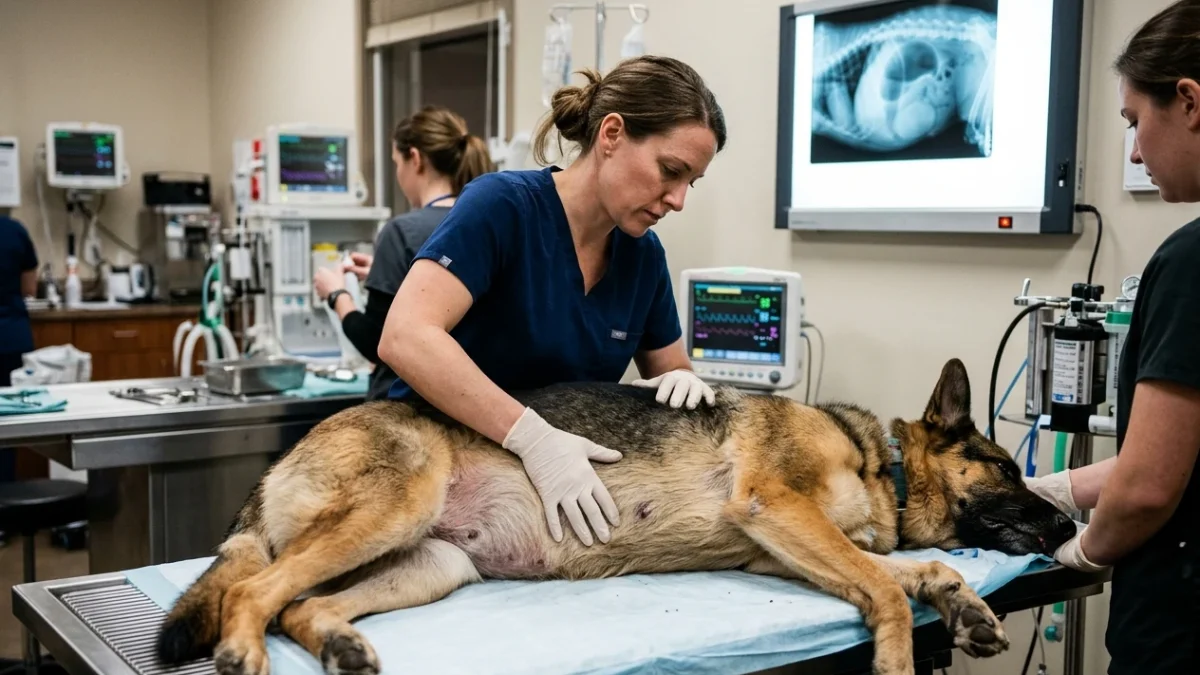
Upon arrival at a veterinary emergency facility, the medical team will initiate simultaneous stabilization and diagnostic protocols. Two large-bore intravenous catheters are typically placed in the front legs (cephalic veins) to rapidly deliver aggressive crystalloid and colloid fluid therapy, directly combating the profound hypovolemic shock. Flow-by oxygen therapy or intubation is initiated to address tissue hypoxia.
The definitive diagnosis of GDV is achieved rapidly via abdominal radiography (X-rays), specifically utilizing a right lateral view. This radiograph reveals the classic “double bubble,” “smurf hat,” or “Popeye’s arm” appearance, created by the displaced, gas-filled pylorus situated dorsal and cranial to the fundus, compartmentalized by a soft tissue band of the twisted stomach wall.
Once diagnosed and initially stabilized, the dog is placed under general anesthesia for an emergency exploratory laparotomy (abdominal surgery). The surgical procedure involves three primary, critical objectives: decompression of the stomach via orogastric intubation or trocharization, manual derotation to restore normal anatomical positioning and relieve the vascular obstruction, and a meticulous assessment of tissue viability. The surgeon will meticulously inspect the entire gastric wall and the spleen. If severe necrosis is present, partial gastrectomy (removal of the dead portions of the stomach tissue) or splenectomy (complete removal of the spleen) must be performed to prevent postoperative sepsis.
Prophylactic Gastropexy: Preventing Torsion
The final and most critical step of any GDV surgery, which is also increasingly performed electively as a primary preventive measure, is a gastropexy. This surgical procedure involves permanently suturing the outer layer of the right side of the stomach (specifically the pyloric antrum) to the interior right abdominal body wall. A gastropexy does not prevent gastric dilatation (the stomach can still fill with gas or food), but it physically anchors the stomach to prevent the catastrophic twisting (volvulus) from occurring. For highly predisposed breeds, such as Great Danes or Standard Poodles, prophylactic gastropexy is frequently recommended by veterinary surgeons and is often performed simultaneously with routine spaying or neutering using laparoscopic techniques. Clinical studies indicate that a properly performed prophylactic gastropexy reduces the recurrence rate or initial incidence of the volvulus component of GDV from over 70 percent to less than 5 percent, effectively eliminating the immediate threat to life.
Post-Operative Recovery and Long-Term Prognosis
The first 48 to 72 hours following GDV surgery are intensive and require continuous hospitalization. Patients are closely monitored for life-threatening cardiac arrhythmias, which often peak within this postoperative window. Aggressive pain management, targeted fluid therapy to correct electrolyte imbalances (particularly hypokalemia), and specialized nutritional support are maintained. The overall survival rate for dogs undergoing surgery for GDV ranges from 75 to 85 percent, heavily dependent on the duration of symptoms prior to surgical intervention and the extent of tissue necrosis present at the time of operation. Dogs requiring concurrent splenectomy or partial gastrectomy face statistically lower survival rates and higher risks of postoperative complications, underscoring the absolute necessity of rapid triage.
Differential Diagnosis: Is It Something Else?
While GDV must always be assumed and treated as the primary differential until definitively proven otherwise via radiography, other severe conditions can present with similar acute symptoms. Acute necrotizing pancreatitis, severe intestinal obstruction (such as foreign body ingestion), or acute hemorrhagic gastroenteritis (AHGE) can cause profound abdominal pain, shock, and retching. For an exhaustive guide on distinguishing various presentations and determining intervention necessity, refer to our comprehensive resource on dog vomiting home monitoring versus emergency red flags. Furthermore, if the primary symptom is severe lethargy and generalized trembling rather than visible abdominal distension or retching, consult our detailed protocol on dog shaking and lethargic differential triage.
Sources and Evidence
The clinical protocols, risk assessments, and surgical descriptions outlined in this article are strictly grounded in established veterinary surgical texts and peer-reviewed literature. For further technical data, refer to the following sources:
- Gastric Dilatation-Volvulus in Dogs (PubMed: PMC5867011)
- Risk factors associated with gastric dilatation-volvulus in dogs (PubMed: 28812613)
- Prophylactic gastropexy in dogs (PubMed: 31278893)
Caregiver Safety Checklist for GDV Prevention
- Identify objectively if your dog belongs to a high-risk deep-chested breed.
- Divide daily food rations into two or three smaller meals rather than feeding one massive meal.
- Utilize specialized slow-feeder bowls or feeding puzzles to prevent rapid consumption and minimize air swallowing.
- Feed from the floor rather than using elevated food bowls, unless specifically directed by a veterinary specialist for an unrelated medical condition (e.g., megaesophagus).
- Enforce a strict rest period of at least one hour before and one to two hours after meals, completely avoiding vigorous exercise or active play.
- Familiarize yourself precisely with the exact location, phone number, and operating hours of the nearest 24-hour veterinary emergency facility.
- Discuss the option and optimal timing of a prophylactic gastropexy with your primary care veterinarian if you own an at-risk breed.
Source transparency
Reference mix for this guide
Source labels describe the type of organization behind each reference; they are not a claim that any outside organization reviewed this article.
- dog vomiting home monitoring versus emergency red flags Reference source · newspet.net
- dog shaking and lethargic differential triage Reference source · newspet.net
- Gastric Dilatation-Volvulus in Dogs (PubMed: PMC5867011) Government or public agency · pubmed.ncbi.nlm.nih.gov
- Risk factors associated with gastric dilatation-volvulus in dogs (PubMed: 28812613) Government or public agency · pubmed.ncbi.nlm.nih.gov
- Prophylactic gastropexy in dogs (PubMed: 31278893) Government or public agency · pubmed.ncbi.nlm.nih.gov