This guide is general education for pet owners. If your pet has urgent symptoms, possible poisoning, injury, breathing trouble, collapse, severe pain, or sudden decline, contact a licensed veterinarian or local emergency service.
Dosages must be calculated by a licensed veterinarian based on species, weight, and bloodwork. Never administer human medications or self-adjust prescription doses for pets.
Editorial standard
How this guide was prepared
Feline jaundice, clinically termed icterus, is an unmistakable physical sign characterized by vivid yellowish pigmentation of the mucous membranes, sclera (the white of the eyes), inner ear flaps (pinnae), and skin. Unlike mild skin conditions, jaundice is never a primary disease entity; it is a critical physiological manifestation of hyperbilirubinemia—an abnormal accumulation of yellow bile pigments in circulating blood exceeding 2.0 mg/dL (34 μmol/L). In felines, unique anatomical features (including a shared common bile duct and pancreatic duct) predispose cats to severe hepatic and biliary inflammation. Recognizing early subtle yellowing and understanding the diagnostic stratification between pre-hepatic, hepatic, and post-hepatic causes is vital for executing life-saving clinical triage.
Quick Answer: What Causes Cat Jaundice?
Yellowing of a cat’s skin, ears, or eyes indicates that bilirubin is building up in the bloodstream. In cats, over 80 percent of cases stem from three life-threatening conditions: acute hepatic lipidosis (fatty liver disease from not eating for over 48 hours), feline triaditis / cholangiohepatitis (inflammation of the bile ducts, liver, and pancreas), or immune-mediated hemolytic anemia (massive red blood cell destruction). Home observation is dangerous; prompt veterinary blood panels, abdominal ultrasound, and supportive fluid and nutritional therapy are required.
Anatomical & Physiological Mechanisms: The Three Tiers of Icterus
Bilirubin is a natural metabolic breakdown product of hemoglobin released during the routine clearance of senescent red blood cells. Veterinary clinicians stratify feline jaundice into three distinct anatomical categories:
- Pre-Hepatic (Hemolytic Icterus): Occurs when red blood cells are destroyed faster than the liver can conjugate and excrete bilirubin. Common triggers include infectious hemoplasmosis (Mycoplasma haemofelis), immune-mediated hemolytic anemia (IMHA), neonatal isoerythrolysis, or severe toxic ingestion (e.g., zinc or paracetamol/acetaminophen). The cat presents with ghost-pale gums that subsequently turn yellow, accompanied by rapid breathing, bounding pulses, and hemoglobinuria.
- Hepatic (Hepatocellular Icterus): Originates from direct parenchymal hepatocyte dysfunction or intrahepatic cholestasis, preventing the liver from processing and excreting bilirubin. The classic and most frequent feline cause is hepatic lipidosis (fatty liver disease), which occurs when an overweight cat stops eating for 2 to 7 days, causing massive peripheral triglyceride deposition that overwhelms hepatocytes. Other etiologies include suppurative or lymphocytic cholangiohepatitis, toxic hepatopathy, lymphoma, and dry or wet Feline Infectious Peritonitis (FIP).
- Post-Hepatic (Obstructive Icterus): Results from physical obstruction of the extrahepatic biliary system, preventing bile from flowing from the gallbladder into the duodenum. Common causes include cholelithiasis (gallstones), biliary sludge (inspissated bile), acute pancreatitis compressing the common bile duct, and biliary adenocarcinomas. The common bile duct and pancreatic duct fuse into a single channel before entering the feline duodenum, explaining why feline pancreatitis, cholangitis, and inflammatory bowel disease frequently occur simultaneously in a syndrome termed “triaditis.”
Clinical Emergency Triage Matrix
| Triage Tier | Clinical Presentation | Required Action |
|---|---|---|
| Tier 1: Immediate Emergency (0-2h) | Obvious bright yellow pigmentation of sclera, gums, and skin; acute collapse, profound weakness, or hypothermia (<37.0°C); neurologic signs (head pressing, disorientation, seizures indicating hepatic encephalopathy); protracted vomiting of dark bile or blood; severe respiratory distress. | Transport immediately to a 24-hour veterinary emergency hospital. High risk of fulminant liver failure, septic peritonitis, or severe hemolytic crisis requiring emergency blood transfusion or surgical biliary decompression. |
| Tier 2: Same-Day Veterinary Evaluation (12-24h) | Subtle yellow hue on the inner ear flaps or hard palate; complete anorexia exceeding 24–48 hours; progressive weight loss with vomiting; dark orange-brown urine; enlarged, distended abdomen with palpable fluid wave (suspected FIP effusion). | Schedule an urgent same-day veterinary appointment. Immediate complete blood count (CBC), serum biochemistry (ALT, AST, ALP, GGT, total bilirubin), and abdominal ultrasonography are mandatory. |
| Tier 3: Early Clinical Investigation | Cat is eating normally and active, but urine appears noticeably darker amber; slight elevation in liver enzymes detected on routine geriatric wellness screening without outward clinical illness. | Schedule follow-up diagnostics within 48–72 hours for pre-and-post-prandial bile acids testing and baseline abdominal imaging. |
Dangerous Home Pitfalls: What NOT to Do (Rule S1)
- NEVER assume yellowing will resolve with home rest: Bilirubin accumulation reflects critical organ failure. Waiting even 24 to 48 hours can allow acute hepatic lipidosis to progress to irreversible intrahepatic cholestasis and multi-organ failure.
- NEVER force-feed pureed food by oral syringe: Force-feeding a nauseated, jaundiced cat carries a severe risk of aspiration pneumonia and creates permanent learned food aversion. Safe feeding requires in-hospital placement of an esophageal (E-tube) or nasogastric feeding tube under veterinary care.
- NEVER administer human supplements, milk thistle tinctures, or herbal detoxes: Many human liquid tinctures contain alcohol or unstandardized herbal extracts that exacerbate feline liver failure. Furthermore, cats have defective hepatic glucuronidation pathways, making them hypersensitive to herbal toxicities.
- NEVER administer acetaminophen (Tylenol) under any circumstance: Acetaminophen is rapidly fatal to cats; it oxidizes hemoglobin into methemoglobin and induces massive hepatocellular necrosis within single milligrams.
Veterinary Diagnostic Workup & Clinical Management
Differentiating the underlying cause requires systematic laboratory and diagnostic imaging protocols:
Complete Blood Count & Blood Smear: Evaluates for severe regenerative or non-regenerative anemia. Manual slide review under oil immersion identifies Mycoplasma haemofelis epicellular organisms, spherocytes (hallmark of immune-mediated destruction), or Heinz bodies (oxidative damage).
Comprehensive Serum Biochemistry: Evaluates hepatocellular leakage enzymes (ALT, AST) versus cholestatic enzymes (ALP, GGT). In hepatic lipidosis, alkaline phosphatase (ALP) is massively elevated, while gamma-glutamyl transferase (GGT) remains normal or only mildly elevated—a diagnostic dissociation characteristic of fatty liver. Concurrent severe GGT elevation suggests cholangitis or extrahepatic biliary duct obstruction.
Abdominal Ultrasonography: High-resolution ultrasound evaluates liver echogenicity (diffusely hyperechoic in lipidosis), gallbladder wall thickness, biliary duct diameter (tortuous dilation indicates physical obstruction), pancreatic swelling, and the presence of peritoneal effusion (characteristic of wet FIP or septic peritonitis).
Intensive Enteral Nutrition: In feline hepatic lipidosis, nutritional therapy is the primary definitive cure. Once the cat is stabilized with balanced IV fluids and antiemetics, an esophagostomy feeding tube (E-tube) is placed under light anesthesia. This allows caregivers to administer high-protein, calorie-dense veterinary therapeutic diets directly into the esophagus for several weeks until natural appetite returns.
Frequently Asked Questions
Where is the easiest place to check for jaundice in a cat?
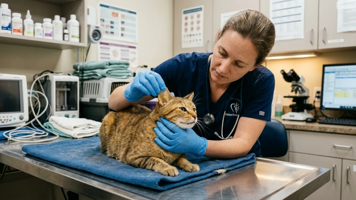
The clearest anatomical locations to check for early yellow discoloration are the inner surface of the ear flaps (pinnae), the white sclera surrounding the pupils, the gums, and the hard palate on the roof of the mouth. In cats with dark fur or pigmented gums, the ear flaps and whites of the eyes provide the most reliable indicators.
Can a cat survive jaundice?
Yes. The prognosis depends entirely on the underlying etiology and how quickly treatment begins. Hepatic lipidosis carries an 80 to 90 percent recovery rate with early in-hospital feeding tube placement. Conversely, untreated liver failure, complete extrahepatic biliary obstructions, or advanced FIP carry guarded prognoses without specialized emergency care.
What does a cat’s urine look like when they have jaundice?
When circulating conjugated bilirubin spills into the urine through the renal tubules (bilirubinuria), the cat’s urine changes from normal pale yellow to a deep, dark orange, tea-colored, or brown shade that noticeably stains litter box clumps. Bilirubin in feline urine is always an abnormal finding requiring veterinary evaluation.
Scientific Sources and Literature
- Valtolina S, et al. “Feline hepatic lipidosis: pathophysiology, diagnostic approach, and intensive management.” Journal of Feline Medicine and Surgery. PMID: 32185842
- Forman MA, et al. “Feline Pancreatitis and Triaditis: A Review of the Pathophysiology and Clinical Signs.” Journal of Veterinary Internal Medicine. PMID: 23812836
- Sparkes AH, et al. “Consensus Guidelines on Feline Systemic and Hepatic Infectious Disorders.” Journal of Feline Medicine and Surgery. PMID: 27151044
Caregiver Jaundice Assessment Checklist
- Inspect your cat’s inner ear flaps and eye sclera under bright, direct natural sunlight for any subtle yellow tint.
- Monitor food intake daily; never allow an overweight cat to go without food for more than 24 to 48 hours.
- Observe litter box clumps for dark amber, tea-colored, or orange-stained urine.
- Document any episodes of vomiting, noting frequency and the presence of yellow or green bile.
- Check for abdominal enlargement or swelling that might indicate fluid accumulation (ascites).
- Contact an emergency veterinary hospital immediately upon observing yellowing; do not attempt home observation.
Source transparency
Reference mix for this guide
Source labels describe the type of organization behind each reference; they are not a claim that any outside organization reviewed this article.
- Cat Not Eating & Anorexia Timeline Reference source · newspet.net
- Cat Vomiting: Acute vs Chronic Differentiation Reference source · newspet.net
- Cat Drinking Lots of Water: Diabetes vs CKD Reference source · newspet.net
- Cat Drooling & Oral Stomatitis Guide Reference source · newspet.net
- PMID: 32185842 Government or public agency · pubmed.ncbi.nlm.nih.gov
- PMID: 23812836 Government or public agency · pubmed.ncbi.nlm.nih.gov
- PMID: 27151044 Government or public agency · pubmed.ncbi.nlm.nih.gov