This guide is general education for pet owners. If your pet has urgent symptoms, possible poisoning, injury, breathing trouble, collapse, severe pain, or sudden decline, contact a licensed veterinarian or local emergency service.
Dosages must be calculated by a licensed veterinarian based on species, weight, and bloodwork. Never administer human medications or self-adjust prescription doses for pets.
Editorial standard
How this guide was prepared
The first ten to fourteen days following canine surgery represent a critical physiological window where wound tensile strength is entirely dependent on suture integrity and delicate cellular fibrin scaffolding. Whether recovering from routine elective procedures (such as ovariohysterectomy or castration) or complex emergency interventions (such as exploratory laparotomy or orthopedic stabilization), the surgical incision requires vigilant daily monitoring. In veterinary recovery, complications range from benign, self-limiting fluid accumulations to catastrophic full-thickness abdominal wall dehiscence with intestinal evisceration. Differentiating between normal inflammatory healing, seromas, bacterial abscesses, and surgical emergencies is essential for preventing septic peritonitis and irreversible tissue trauma.
Quick Answer: Normal Incision vs. Complication
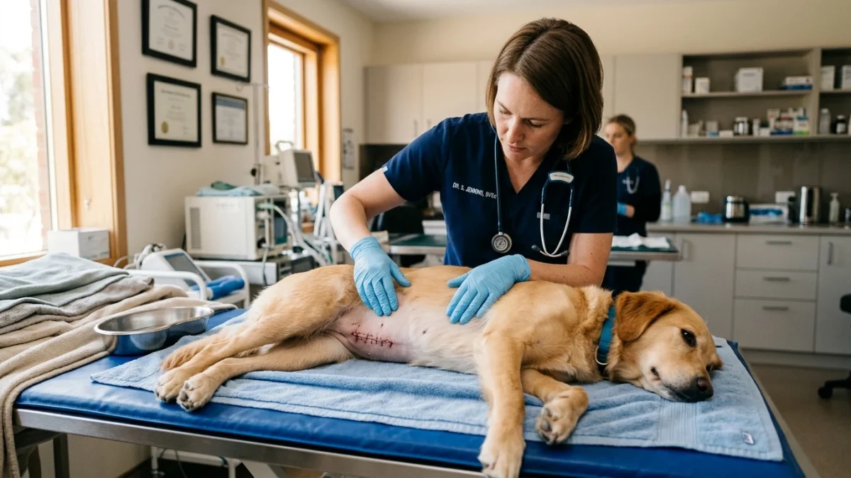
A normal healing surgical incision shows edges neatly held together with minimal swelling, slight redness that fades after 48 hours, and perhaps a small droplet of clear or blood-tinged fluid within the first 24 hours. A seroma presents as a soft, squishy, non-painful swelling beneath the skin without heat or foul odor. An infected abscess is hot, painful, firm, and discharges opaque yellow or green pus. Full-thickness dehiscence—an open gap revealing subcutaneous tissue or internal organs—requires immediate emergency surgical revision.
The Cellular Stages of Incision Healing
Incision repair follows four coordinated physiological phases. Understanding this biological progression helps caregivers spot pathological deviations early:
- Hemostasis and Acute Inflammation (Days 1–4): Immediately following skin apposition, platelets form a fibrin clot to seal the capillary beds. Neutrophils and macrophages migrate to the site to engulf debris and bacteria. Mild localized erythema (redness) and minor swelling are expected physiological responses to surgical trauma and suture reactivity. Tensile strength is near zero; the wound relies completely on mechanical suture holds.
- Proliferation and Granulation (Days 4–14): Fibroblasts synthesize collagen, forming vascular granulation tissue. New capillaries bridge the incision margin (angiogenesis), and epithelial cells migrate across the surface to restore skin barrier integrity. A firm, linear healing ridge typically develops along the suture line by Day 5; this firm ridge is a healthy sign of collagen deposition, not an infection.
- Maturation and Remodeling (Day 14 through Months): Suture materials are absorbed or removed. Disorganized Type III collagen is gradually replaced by cross-linked Type I collagen fibers, restoring up to 80 percent of original uninjured dermal tensile strength over several months.
Clinical Emergency Triage Matrix
| Triage Tier | Clinical Presentation | Required Action |
|---|---|---|
| Tier 1: Immediate Emergency (0-2h) | Complete gap in the incision with protruding internal tissue (subcutaneous fat or intestine/evisceration); active arterial or heavy venous bleeding that soaks through towels; sudden pale gums, hypothermia, collapse, or shock following abdominal surgery. | Cover the incision loosely with a clean, moist towel (saline or clean water). Do not apply pressure or manipulate tissue. Transport immediately to an emergency veterinary hospital for emergency surgical re-closure. |
| Tier 2: Same-Day Veterinary Evaluation (12-24h) | Incision margins separating without visible deep tissue; thick yellow or green purulent discharge with foul odor; rapidly enlarging swelling that feels hot and painful to the touch; fever (>39.5°C) accompanied by complete anorexia and lethargy. | Contact your primary surgeon or emergency clinic for a same-day evaluation. Requires wound culture, sterile debridement, and targeted systemic antimicrobial therapy. |
| Tier 3: Home Monitoring & Strict Rest | Soft, painless, fluid-filled swelling (seroma) directly under the incision without redness, heat, or discharge; incision edges tightly apposed; dog is bright, eating normally, and pain is well-controlled. | Enforce strict crate confinement and short leash-only elimination walks. Apply cold compresses (wrapped in a clean cloth) for 10 minutes twice daily if approved by your surgeon. Never attempt to drain fluid at home. |
Dangerous Home Pitfalls: What NOT to Do (Rule S1)
- NEVER apply hydrogen peroxide, rubbing alcohol, or witch hazel: These harsh chemicals are directly cytotoxic to proliferating fibroblasts and newly formed capillary buds. Pouring peroxide on a surgical wound dissolves delicate granulation tissue, delays wound closure, and dramatically increases infection risks.
- NEVER apply human topical antibiotic ointments (Neosporin, Polysporin): Topical ointments keep surgical incisions excessively moist, causing maceration of dermal edges. Furthermore, if the dog licks the ointment, active ingredients like bacitracin or polymyxin B can cause acute toxic gastroenteritis.
- NEVER attempt to drain a seroma with a home needle: A seroma is a sterile fluid pocket. Inserting an unsterile home needle introduces aggressive skin bacteria directly into the deep tissue plane, instantly transforming a harmless seroma into a life-threatening deep subcutaneous abscess.
- NEVER remove the Elizabethan collar (cone) unattended: A dog’s tongue can completely tear out surgical staples and rip open abdominal incisions in under 60 seconds. The cone must remain secured 24/7 until your veterinarian removes sutures and formally clears the patient.
Differentiating Seroma vs. Abscess vs. Hernia
Caregivers frequently panic when a lump appears near an incision. Three distinct entities account for post-operative swelling:
1. Post-Surgical Seroma: A seroma is an accumulation of sterile lymphatic fluid and serum in dead space created between tissue layers during surgery. It is heavily associated with premature physical activity, running, or jumping post-surgery. Clinically, a seroma feels like a soft, squishy water balloon beneath the skin. It is cool to the touch, non-painful on gentle palpation, and the overlying skin remains normal in color. The vast majority of seromas reabsorb naturally over 10 to 21 days with strict cage rest.
2. Surgical Site Infection & Abscess: An abscess occurs when bacteria (predominantly Staphylococcus pseudintermedius or Streptococcus) colonize the surgical site, often introduced by the dog licking the wound. Unlike a seroma, an abscess is firm, hot, exquisitely tender, and accompanied by surrounding tissue erythema. As neutrophils liquefy, purulent exudate (pus) builds pressure, eventually weeping between suture points or requiring surgical drainage and systemic antibiotics.
3. Incisional Hernia: If the deep muscular holding layer (the linea alba in abdominal surgery) fails while the superficial skin remains intact, abdominal contents (falciform fat, omentum, or loops of bowel) herniate into the subcutaneous space. A hernia often feels soft and can sometimes be gently reduced (pushed back into the abdomen). If a loop of intestine becomes incarcerated or strangulated within the hernia ring, blood supply is cut off, precipitating acute vomiting, severe abdominal pain, and rapid endotoxic shock requiring emergency surgery.
Frequently Asked Questions
What does a normal healing surgical incision look like on a dog?
A normal incision has clean, closely apposed skin edges held securely by sutures, staples, or surgical glue. Minor redness and slight bruising around the margins are normal for the first 48 hours. By Day 5 to 7, a firm, non-painful linear ridge should be palpable along the scar line, indicating active collagen remodeling.
Can a dog sleep without an Elizabethan collar (cone) after surgery?
No. Most surgical site dehiscing occurs at night when owners are asleep. An unattended dog can lick through sutures in minutes, introducing oral bacteria and ripping open deep muscle layers. The collar must stay on continuously until suture removal.
What should I do if my dog’s surgical incision opens up?
If the incision gaps open, cover it immediately with a clean cloth moistened with warm saline or clean water to protect exposed tissues from drying and contamination. Prevent your dog from touching the area, keep them calm and strictly confined, and transport them immediately to an emergency veterinary clinic for surgical repair.
Scientific Sources and Literature
- Weese JS. “Investigation of antimicrobial prophylaxis in small animal surgery.” Veterinary Surgery. PMID: 28844883
- Rosenthal K. “Postoperative complications in small animal soft tissue surgery: seroma, dehiscence, and infection.” Journal of the American Animal Hospital Association. PMID: 25146654
- Gastropexy and Laparotomy Wound Management in Canines. Journal of Veterinary Internal Medicine. PMID: 31278893
Caregiver Post-Surgical Incision Checklist
- Inspect the incision twice daily under bright direct lighting; check for swelling, redness, edge separation, or discharge.
- Keep the Elizabethan collar securely fastened 24/7 until sutures are removed by your veterinarian.
- Enforce strict cage rest; allow elimination trips outside only on a short handheld leash, preventing all running, jumping, and stairs.
- Keep the incision completely clean and dry; do not bathe the dog, allow swimming, or apply any unprescribed creams or ointments.
- Administer all prescribed veterinary pain medications and anti-inflammatories strictly on schedule to prevent anxiety and self-trauma.
- Contact your veterinary surgeon immediately if you notice foul odors, oozing pus, sudden swelling, or visible gaps between stitches.
Source transparency
Reference mix for this guide
Source labels describe the type of organization behind each reference; they are not a claim that any outside organization reviewed this article.
- Dog Bleeding & Hemorrhage First Aid Reference source · newspet.net
- Dog Limping & Orthopedic Triage Reference source · newspet.net
- Dog Bloat (GDV) Emergency Signs Reference source · newspet.net
- Dog Shaking & Lethargic Differential Triage Reference source · newspet.net
- PMID: 28844883 Government or public agency · pubmed.ncbi.nlm.nih.gov
- PMID: 25146654 Government or public agency · pubmed.ncbi.nlm.nih.gov
- PMID: 31278893 Government or public agency · pubmed.ncbi.nlm.nih.gov