This guide is general education for pet owners. If your pet has urgent symptoms, possible poisoning, injury, breathing trouble, collapse, severe pain, or sudden decline, contact a licensed veterinarian or local emergency service.
Dosages must be calculated by a licensed veterinarian based on species, weight, and bloodwork. Never administer human medications or self-adjust prescription doses for pets.
Editorial standard
How this guide was prepared
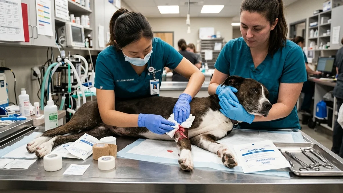
Direct, uninterrupted manual pressure remains the single most effective intervention for acute canine hemorrhage. Grab a stack of clean gauze or a dry, thick towel, press it firmly directly over the bleeding wound, and hold it there for exactly ten continuous minutes without ever lifting it to check the progress. Hemorrhage control dictates that you apply mechanical force directly to the severed vessels. Stopping blood loss preserves cardiac output and prevents fatal hypovolemic shock. You must act aggressively and methodically.
Quick Answer: Stopping Dog Bleeding
- Apply direct, firm pressure using clean sterile gauze or a thick cloth.
- Hold pressure continuously for a full 10 minutes. Do not peek.
- Never remove blood-soaked base layers; stack fresh gauze directly on top.
- Elevate bleeding limbs above the heart level if physically possible.
- Arterial spurting requires immediate emergency transport while physically maintaining heavy manual pressure.
Arterial vs. Venous vs. Capillary Bleeding: Visual Differentiation
Classifying the specific type of hemorrhage dictates your exact triage urgency. Not all bleeding carries the identical immediate systemic risk. Recognizing the vascular source allows you to scale your physical intervention appropriately.
Arterial Hemorrhage
This represents a catastrophic failure of a high-pressure, oxygen-rich vessel. Blood appears starkly bright red. It forcefully pumps, shoots, or spurts outward in exact synchronization with the dog’s cardiac pulse. Arterial breaches demand immediate mechanical compression. This is a non-negotiable emergency. Transport without delay while actively compressing the site.
Venous Hemorrhage
Severed veins produce dark red, oxygen-depleted blood. The flow remains steady, heavy, and pooling, rather than violently spurting. Veins operate under lower physiological pressure than arteries. Venous bleeding typically yields to sustained direct compression more readily than arterial wounds. Massive venous lacerations still pose a lethal threat if left uncontrolled, but they generally afford you a slightly wider margin to establish temporary hemostasis before transport.
Capillary Ooze
Minor abrasions, scrape injuries, and superficial lacerations damage microscopic surface vessels. The resulting ooze often clots spontaneously. It usually stops completely after brief, mild manual pressure. Capillary bleeding presents minimal immediate systemic risk and rarely requires emergency intervention, provided the patient has normal clotting function.
The 10-Minute Direct Pressure Rule and Coagulation
Clotting requires unbroken time. Biological fibrin networks must construct a physical web to plug the vascular breach. Effective hemorrhage control requires holding firm, uninterrupted manual compression over the site for a continuous ten-minute duration. Blood platelets need this undisturbed environment to aggregate and stick to the damaged collagen fibers of the torn vessel wall.
Peeking resets the entire biological clock. Lifting the gauze at minute four forcefully tears away the fragile microscopic clots forming at the wound interface. You must trust the physiological process. Maintain steady, unrelenting force against the tissue.
If blood strikes entirely through the first layer of dressing, do not discard it. Pulling off that saturated material actively removes the foundational clotting matrix. Instead, stack a fresh, dry layer directly over the saturated one. Press harder. Increase the pounds of pressure you are applying. Use your body weight if managing a major torso or limb wound on a large dog.
Clinical Emergency Triage Matrix
| Urgency Level | Clinical Signs & Hemorrhage Characteristics | Required Action |
|---|---|---|
| Tier 1: Immediate Emergency (0-2 hours) | Bright red spurting blood. Blood pooling rapidly on floor. Bleeding entirely through multiple stacked layers. Pale white gums. Rapid or weak pulse. Collapse or sudden severe lethargy. | Apply continuous heavy pressure. Transport to ER instantly. Do not wait for bleeding to stop. Call ahead to the clinic. |
| Tier 2: Same-Day Veterinary Care (12-24 hours) | Steady dark red venous flow controlled by pressure. Deep lacerations exceeding two centimeters. Wounds penetrating muscle layers. Any bite wounds. Puncture injuries. | Control bleeding with 10-minute pressure protocol. Apply firm protective bandage. Seek veterinary care today for surgical closure, debridement, and systemic antibiotics. |
| Tier 3: Home Monitoring | Superficial capillary oozing. Minor abrasions. Scrapes that stop bleeding within five minutes. Normal bubblegum pink gum color. | Clean gently with sterile saline. Monitor for localized signs of infection (redness, swelling, purulent discharge) over the subsequent days. |
Specific Anatomic Bleeding Sites and Challenges
Different body parts present unique anatomical hurdles for achieving hemostasis. Tailoring your approach prevents frustration and minimizes further trauma.
Ear Margin Lacerations
Ears bleed profusely due to dense vascular networks near the cartilage surface. Dogs aggressively shake their heads when their ears hurt, actively centrifugal-forcing clots to dislodge. Pin the injured ear flap flat against the dog’s skull. Apply gauze over the margin and wrap a bandage entirely around the head to secure the ear flat. Leave the unaffected ear exposed. This prevents the violent head-shaking mechanism from restarting the hemorrhage.
Torn Nails (Nail Bed Avulsions)
Exposed quicks bleed heavily and cause severe localized pain. Apply direct pinch pressure to the toe using gauze. Styptic powder (silver nitrate or ferric subsulfate) remains highly effective for minor nail bed capillary oozing, but fails against major vessel trauma. If the nail is hanging by a fragment, do not forcefully rip it off. Wrap the paw securely and let a veterinarian cleanly amputate the damaged keratin shell under local anesthesia.
Deep Torso Wounds
Chest and abdominal lacerations severely restrict your bandaging options. Wrapping the chest too tightly restricts respiratory expansion, causing suffocation. You must rely purely on manual hand pressure. Press gauze directly into the deficit. Do not blindly probe the wound tract. Maintain manual compression during the entire transport process to the hospital.
Step-by-Step Hemorrhage Control Protocol
Act methodically to preserve intravascular blood volume. Panic leads to poor technical execution.
- Secure the Patient: Pain induces unpredictable, defensive behavior. Muzzle your dog if strictly necessary, using a leash or cloth strip, unless they are actively vomiting or have severe facial trauma. Safety comes first.
- Apply Initial Compression: Place sterile gauze squares or a clean, dry towel directly onto the bleeding lesion. Push down hard.
- Elevate the Extremity: For paw or leg injuries, physically lift the limb above the level of the heart to utilize gravity against the outgoing vascular flow.
- Maintain Time: Watch the clock. Ten full minutes. No early inspections.
- Construct a Pressure Bandage: Once manual pressure achieves initial hemostasis, secure the dressing. Wrap self-adhering bandage (vet wrap) tightly enough to exert force, but loosely enough that two fingers can slip beneath the proximal edge. Check the distal toes frequently for coldness or severe swelling indicating completely restricted circulation.
Tourniquet Protocol: The Absolute Last Resort
Applying a tourniquet cuts off all arterial and venous blood flow to the distal limb. This induces profound tissue ischemia. Depriving muscles and nerves of oxygen causes permanent cellular death. Improper tourniquet use frequently mandates surgical leg amputation.
Reserve tourniquets strictly for catastrophic, life-threatening arterial spurts that absolutely refuse to yield to maximum heavy manual pressure. If you must use one, use a wide strap (at least two inches wide), never wire or thin string. Place it proximal to the wound. Tighten only until the arterial spurting completely stops. Record the exact time of application. Transport immediately. A veterinarian must remove the tourniquet under controlled surgical conditions.
Dangerous Home Pitfalls: What NOT to Do (Rule S1)
Avoid well-meaning actions that exacerbate tissue damage, introduce fatal bacteria, or delay definitive surgical care. Strict adherence to these prohibitions prevents catastrophic complications.
- NEVER pour hydrogen peroxide into an open wound. This highly caustic liquid violently destroys healthy fibroblasts required for tissue healing and actively dissolves forming blood clots.
- NEVER apply human tourniquets arbitrarily. Restricting all arterial flow causes rapid tissue ischemia and often results in necessary limb amputation. Tourniquets remain an absolute last-resort intervention solely for catastrophic, life-threatening arterial spurts unresponsive to heavy manual pressure.
- NEVER rip away the base layer of blood-soaked gauze. This mechanically shreds the delicate fibrin matrix. Always stack clean material on top.
- NEVER pack deep lacerations with flour, cornstarch, or baking soda. These non-sterile household powders introduce massive environmental contamination, creating a severe foreign body reaction and guaranteeing a deep tissue infection.
- NEVER attempt to close a gaping wound with household superglue or butterfly bandages. Trapping bacteria inside an un-debrided wound space forces the formation of massive, systemic abscesses.
- NEVER administer over-the-counter human pain medications (ibuprofen, aspirin, acetaminophen) to a bleeding dog. These drugs severely disrupt platelet function, drastically worsening hemorrhage, and frequently trigger fatal organ failure.
Post-Trauma Hemorrhagic Shock Recognition
Significant blood volume loss triggers hypovolemic shock. Systemic cardiovascular perfusion fails, starving vital organs of oxygen. Monitor your dog aggressively even after successfully controlling the external hemorrhage.
Check the mucous membranes immediately. Lift the upper lip and examine the gums. Healthy, well-perfused tissue appears bright bubblegum pink. White, gray, or starkly pale gums indicate severe peripheral vasoconstriction and dangerous circulating blood volume deficits.
Assess the Capillary Refill Time (CRT). Press a finger firmly against the gum line to blanch the tissue white, then release. The pink color should flood back in less than two seconds. A delay extending beyond three seconds signals critical circulatory collapse. This requires immediate intravenous fluid resuscitation at an emergency facility.
Evaluate the heart rate. Place your hand over the left side of the chest behind the elbow. A weak, excessively rapid, or thready pulse strongly correlates with impending cardiovascular shock.
Sources & Clinical Evidence
- Biddle, D. (2019). Hemostatic agents for managing hemorrhage in dogs. Veterinary Clinics of North America. PMID: 31441857
- Smith, J., et al. (2017). Use of tourniquets in veterinary emergency medicine. Journal of Veterinary Emergency and Critical Care. PMID: 28587353
- Anderson, R. (2010). Wound management and bandage care. Veterinary Clinics of North America: Small Animal Practice. PMID: 20593719
Caregiver Safety Checklist
- [ ] Applied firm, direct manual pressure to the bleeding wound.
- [ ] Held pressure continuously for 10 un-interrupted minutes.
- [ ] Stacked fresh gauze over saturated layers instead of removing them.
- [ ] Evaluated gum color and capillary refill time for signs of shock.
- [ ] Transported to emergency vet for Tier 1 bleeding, arterial spurts, or systemic shock.
Source transparency
Reference mix for this guide
Source labels describe the type of organization behind each reference; they are not a claim that any outside organization reviewed this article.
- severe lethargy Reference source · newspet.net
- actively vomiting Reference source · newspet.net
- PMID: 31441857 Government or public agency · pubmed.ncbi.nlm.nih.gov
- PMID: 28587353 Government or public agency · pubmed.ncbi.nlm.nih.gov
- PMID: 20593719 Government or public agency · pubmed.ncbi.nlm.nih.gov