This guide is general education for pet owners. If your pet has urgent symptoms, possible poisoning, injury, breathing trouble, collapse, severe pain, or sudden decline, contact a licensed veterinarian or local emergency service.
Dosages must be calculated by a licensed veterinarian based on species, weight, and bloodwork. Never administer human medications or self-adjust prescription doses for pets.
Editorial standard
How this guide was prepared
Tick bites on dogs often appear deceptively benign in the initial hours following attachment. While the physical bite site may show only mild localized erythema, the intracellular pathogens transmitted by ixodid ticks initiate complex hematologic and immune-mediated cascades. In canine clinical practice, three primary tick-borne pathogens dominate emergency admissions: Ehrlichia canis, Babesia canis (and Babesia gibsoni), and Borrelia burgdorferi (the causative agent of Lyme disease). Recognizing the distinct clinical presentations of thrombocytopenia, acute hemolytic anemia, and immune-mediated polyarthritis separates routine outpatient monitoring from life-saving emergency veterinary intervention.
Quick Answer: Recognizing Tick-Borne Emergencies
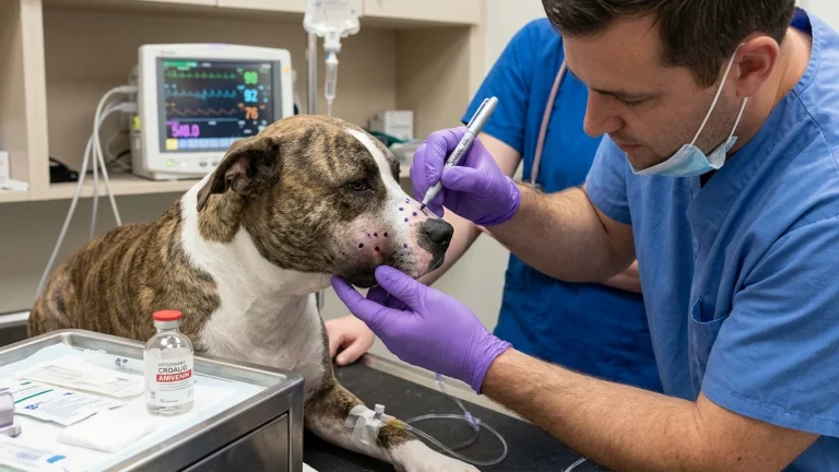
An attached tick that is flat and removed within 24 hours rarely transmits systemic disease. However, if a dog develops an acute fever (>39.5°C / 103.1°F), pale porcelain-white gums, pinpoint red bruises (petechiae) on the mucous membranes or inner ear flaps, dark red urine, or sudden shifting-leg lameness weeks after tick exposure, emergency veterinary diagnostics are required immediately. These signs indicate active platelet destruction, acute red blood cell hemolysis, or septic immune complex deposition.
Pathophysiology: How Three Tick Pathogens Attack Canine Blood
Understanding the targeted cell lines helps caregivers recognize why tick diseases produce such diverse clinical manifestations:
- Ehrlichiosis (Platelet Destruction): Ehrlichia canis is an obligate intracellular rickettsia transmitted primarily by the brown dog tick (Rhipicephalus sanguineus). Once inoculated, the organism infects circulating monocytes, disseminating to the spleen, liver, and lymph nodes. In the acute phase, splenic sequestration and immune-mediated destruction cause severe thrombocytopenia (crashing platelet counts). Without platelets to plug microvascular leaks, dogs develop epistaxis (nosebleeds), petechiae, and intraocular bleeding. Chronic ehrlichiosis causes irreversible bone marrow hypoplasia, shutting down blood cell production entirely.
- Babesiosis (Red Blood Cell Lysis): Babesia species are protozoal hemoparasites transmitted by brown dog ticks and Dermacentor species. The sporozoites penetrate erythrocytes (red blood cells), replicating until the host cells rupture. This creates acute hemolytic anemia. Dogs present with jaundiced (icteric) or ghost-pale mucous membranes, rapid bounding pulse, profound weakness, and hemoglobinuria—dark port-wine or tea-colored urine resulting from massive free hemoglobin filtration through the kidneys.
- Lyme Disease (Immune Complex Polyarthritis & Nephritis): Borrelia burgdorferi is a spirochete transmitted by blacklegged deer ticks (Ixodes scapularis). Rather than directly destroying circulating blood cells, the spirochetes migrate through extracellular connective tissues, triggering intense immune-mediated synovitis (joint inflammation). Weeks to months later, dogs develop acute, hot, swollen joints and shifting lameness. In severe genetic predispositions (notably Golden Retrievers and Labrador Retrievers), circulating antigen-antibody complexes lodge in renal glomeruli, producing acute, rapidly fatal Lyme nephritis.
Clinical Emergency Triage Matrix
| Triage Tier | Clinical Presentation | Required Action |
|---|---|---|
| Tier 1: Immediate Emergency (0-2h) | Active spontaneous nosebleeds (epistaxis); pinpoint bruises (petechiae) or large purple blotches (ecchymoses) on gums, belly, or ears; dark red, brown, or tea-colored urine; white or yellow mucous membranes; acute collapse, profound ataxia, or respiratory distress. | Transport immediately to a 24-hour emergency veterinary hospital. High risk of fatal hemorrhagic shock, acute hemolytic crisis, or renal shutdown requiring emergency packed red blood cell or platelet transfusion. |
| Tier 2: Same-Day Veterinary Care (12-24h) | Fever (>39.5°C) with persistent lethargy; sudden refusal to bear weight on one leg that shifts to another leg the next day; hot, swollen carpal or hock joints; generalized lymph node enlargement (lymphadenomegaly) under the jaw or behind the knees. | Schedule an urgent same-day veterinary appointment for vector-borne diagnostic panels (SNAP 4Dx Plus, PCR, complete blood count, and blood smear cytology). |
| Tier 3: Home Monitoring & Outpatient Care | Single unengorged tick removed cleanly with fine-tipped tweezers; small localized firm bump at the bite site without swelling, redness, or heat; normal appetite, bright demeanor, and active energy. | Disinfect the bite site with chlorhexidine or dilute povidone-iodine. Log the date and geographic location on your calendar. Monitor daily temperature and appetite for the next 30 days. |
Dangerous Home Pitfalls: What NOT to Do (Rule S1)
- NEVER burn an attached tick with matches or lighters: Applying heat causes the tick to regurgitate its salivary contents and stomach fluids directly into your dog’s bloodstream, exponentially multiplying transmission speed.
- NEVER smother ticks with petroleum jelly, alcohol, or nail polish: These folklore methods induce severe tick stress, triggering massive pathogen inoculation before the parasite suffocates.
- NEVER administer human fever reducers (acetaminophen, ibuprofen, naproxen): Human antipyretics cause acute liver necrosis, severe gastric ulceration, and renal papillary necrosis. They do not treat the underlying vector infection and can kill a dog within hours.
- NEVER start leftover antibiotics without definitive testing: Giving random amoxicillin or cephalexin does not eradicate intracellular rickettsia or hemoparasites. It masks symptoms, delays appropriate targeted antiprotozoal or tetracycline therapy, and skews diagnostic PCR testing.
Safe Mechanical Tick Removal: The Veterinary Technique
Safe extraction requires steady mechanical traction rather than chemical manipulation:
- Use Fine-Tipped Tweezers or a Tick Hook: Grasp the tick as close to the dog’s skin surface as physically possible, directly at the mouthparts (capitulum).
- Pull Straight Up with Slow, Steady Force: Do not twist, jerk, or yank with tweezers (tick hooks, conversely, utilize a gentle rotating motion). Jerking tweezers shears the body away, leaving the barbed mouthparts embedded in the dermal layer.
- Manage Retained Mouthparts Calmly: If the microscopic mouthparts snap off in the skin, do not dig aggressively with needles. The dog’s immune system will naturally wall off and expel the non-infectious chitin fragment like a tiny splinter. Disinfect the skin and monitor for secondary bacterial pustules.
- Preserve the Specimen for Identification: Place the removed tick into a sealed plastic bag or small vial containing 70% isopropyl alcohol. Label it with the date. If clinical signs emerge weeks later, providing the physical tick helps your veterinarian identify the precise vector species.
Diagnostic Approach and Veterinary Therapeutics
Definitive diagnosis requires a multi-tiered laboratory strategy. In the veterinary clinic, point-of-care serological tests (such as enzyme-linked immunosorbent assay SNAP tests) detect antibodies to E. canis, B. burgdorferi, and Anaplasma species. However, serology indicates past exposure and antibody response, which may take 2 to 4 weeks to develop post-bite, or may persist long after clinical recovery.
To confirm active acute infection, clinicians perform a Complete Blood Count (CBC) with manual blood smear evaluation under 100x oil immersion. Smear cytology can reveal characteristic Ehrlichia morulae inside monocytes or piroplasms inside erythrocytes during high-parasitemia Babesia crises. Quantitative Polymerase Chain Reaction (PCR) assays provide definitive pathogen DNA identification, distinguishing between species such as Babesia canis and the more drug-resistant Babesia gibsoni.
Therapeutic management depends strictly on the confirmed organism. Rickettsial infections (Ehrlichia, Anaplasma) and spirochetes (Borrelia) respond to targeted prescription antibiotic courses administered over 28 continuous days to prevent chronic persistent carrier states. Babesiosis, being a protozoan parasite, requires specialized antiprotozoal injections (such as imidocarb dipropionate or combination atovaquone and azithromycin). Severe cases presenting with packed cell volumes (PCV) below 15% or platelets under 20,000/μL mandate immediate blood transfusion therapy and intensive hospitalization.
Frequently Asked Questions
How long must a tick stay attached to transmit Lyme or Ehrlichia to a dog?
Transmission times vary significantly by pathogen. Borrelia burgdorferi (Lyme) generally requires 24 to 48 hours of continuous attachment as the spirochetes activate inside the tick’s gut and migrate to salivary glands. In contrast, Ehrlichia canis and Babesia can be transmitted in as few as 3 to 24 hours. Daily manual tick checks remain the most effective defense.
Can tick-borne diseases in dogs be cured completely?
When diagnosed and treated early in the acute phase, dogs typically make a full recovery. However, delayed intervention can result in chronic subclinical carrier states where pathogens sequester in the spleen or bone marrow. Chronic ehrlichiosis or persistent Lyme nephritis carries a guarded long-term prognosis due to irreversible renal or hematopoietic damage.
Why is my dog’s urine dark brown or red after a tick bite?
Dark red, brown, or tea-colored urine following tick exposure is a critical emergency indicator of Babesia infection. The hemoparasites rupture red blood cells en masse (hemolysis), releasing hemoglobin into the bloodstream which filters into the urine (hemoglobinuria). This demands immediate emergency veterinary hospitalization.
Scientific Sources and Literature
- Sainz A, et al. “Guidance for the diagnosis, prevention and treatment of tick-borne diseases in dogs.” Journal of Small Animal Practice. PMID: 25176161
- Solano-Gallego L, et al. “A review of canine babesiosis: the European perspective.” Parasites & Vectors. PMID: 30454794
- Mylonakis ME, et al. “Canine monocytic ehrlichiosis: an update on diagnosis and treatment.” Veterinary Clinics of North America: Small Animal Practice. PMID: 29519128
Caregiver Tick Safety Checklist
- Perform full-body manual tick checks daily, focusing on ear crevices, under the collar, armpits, between toes, and around the groin.
- Extract attached ticks immediately using fine-tipped tweezers pulled straight upward without twisting or crushing.
- Preserve the removed tick in a sealed container of rubbing alcohol with the date noted for veterinary identification.
- Check gum color daily for 30 days post-bite; gums should remain healthy salmon-pink, never pale white or yellow.
- Inspect the abdomen and oral mucous membranes for tiny pinpoint red bruises (petechiae).
- Maintain year-round, veterinarian-recommended isoxazoline oral chews or topical vector preventative therapies.
Source transparency
Reference mix for this guide
Source labels describe the type of organization behind each reference; they are not a claim that any outside organization reviewed this article.
- Dog Shaking & Lethargic Differential Triage Reference source · newspet.net
- Dog Bleeding & Hemorrhage First Aid Reference source · newspet.net
- Dog Limping & Sudden Lameness Triage Reference source · newspet.net
- Dog Blood in Urine (Hematuria) Differential Reference source · newspet.net
- PMID: 25176161 Government or public agency · pubmed.ncbi.nlm.nih.gov
- PMID: 30454794 Government or public agency · pubmed.ncbi.nlm.nih.gov
- PMID: 29519128 Government or public agency · pubmed.ncbi.nlm.nih.gov