This guide is general education for pet owners. If your pet has urgent symptoms, possible poisoning, injury, breathing trouble, collapse, severe pain, or sudden decline, contact a licensed veterinarian or local emergency service.
Dosages must be calculated by a licensed veterinarian based on species, weight, and bloodwork. Never administer human medications or self-adjust prescription doses for pets.
Editorial standard
How this guide was prepared
Canine Intervertebral Disc Disease (IVDD) is the most common cause of acute spinal cord compression, paraparesis, and paralysis in dogs. Driven by a retrocopy insertion of the fibroblast growth factor 4 gene on chromosome 12 (FGF4L2), chondrodystrophic breeds—including Dachshunds (where lifetime prevalence approaches 20 to 25 percent), French Bulldogs, Pembroke Welsh Corgis, Bassett Hounds, Shih Tzus, and Beagles—undergo premature chondroid metaplasia and calcification of the gelatinous nucleus pulposus beginning before 1 year of age. When the dorsal annulus fibrosus ruptures (Hansen Type I extrusion, most frequently between T11 and L3), calcified disc material explodes dorsally into the vertebral canal at high velocity, inflicting both acute contusive concussive injury and sustained mechanical compression on the spinal cord.
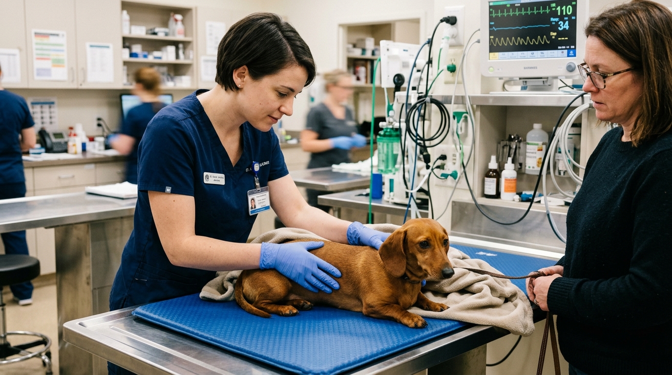
Clinical Quick Answer & Immediate Stabilization Protocol
Primary Clinical Directive for Canine Thoracolumbar & Cervical Intervertebral Disc Disease (Hansen Type I & Type II IVDD): Prognosis and surgical urgency in canine thoracolumbar IVDD depend entirely on the 5-Tier Modified Frankel Neurological Grade and the presence or absence of Conscious Deep Pain Perception (nociception): Dogs in Grade 1 (back pain only) or Grade 2 (ambulatory paraparesis—wobbly walking) can often be managed conservatively with 4 to 6 weeks of strict 24/7 crate confinement and multimodal analgesia, though progressive Grade 2 benefits from decompressive surgery. Once a dog becomes non-ambulatory (Grade 3: cannot stand but can move legs; Grade 4: paralyzed with zero leg movement and urinary retention, but Deep Pain Positive), surgical hemilaminectomy following MRI or CT carries a 90 to 95 percent recovery rate. However, if a paralyzed dog loses Conscious Deep Pain Perception (Grade 5: Deep Pain Negative), recovery drops to ~50 to 60 percent even if emergency hemilaminectomy is performed within 12 to 24 hours, and carries a 10 to 15 percent risk of fatal ascending/descending hemorrhagic myelomalacia.
FGF4Chondroid Metaplasia, Contusion vs. Compression & Deep Pain vs. Withdrawal Reflex
In a normal non-chondrodystrophic juvenile intervertebral disc, the central nucleus pulposus is a hydrated, proteoglycan-rich hydrogel that absorbs axial shock seamlessly. In chondrodystrophic dogs carrying the chromosome 12 FGF4 retrogene, water-binding glycosaminoglycans are rapidly replaced by hyaline cartilage matrix that mineralizes into brittle, chalky material by 12 to 24 months of age. In Hansen Type I extrusion, a sudden torsional twist or vertical impact tears the weakened dorsal annulus fibrosus, ejecting calcified nucleus pulposus into the epidural space. This differs fundamentally from Hansen Type II protrusion—seen in older large-breed dogs such as German Shepherds and Labradors—where fibroid degeneration causes a slow, chronic dome-like bulging of the intact annulus fibrosus over months to years.
Because spinal cord nerve fibers are arranged anatomically from superficial to deep in strict order of diameter and myelination, neurological deficits always progress in a predictable sequence as compression intensifies: (1) first lost are the large, heavily myelinated superficial proprioceptive fibers (causing paw knuckling and drunken ataxia—Grade 2), (2) next lost are the intermediate motor fibers (causing non-ambulatory paresis—Grade 3, and then complete plegia with loss of voluntary urination—Grade 4), and (3) last lost are the tiny, unmyelinated, deeply situated C-fibers and A-delta fibers that transmit Conscious Deep Pain (nociception) bilaterally up the spinothalamic tract to the brain (Grade 5). Because losing deep pain fibers proves that destructive ischemia has spanned the entire cross-section of the spinal cord, Grade 5 status is a time-critical neurosurgical emergency.
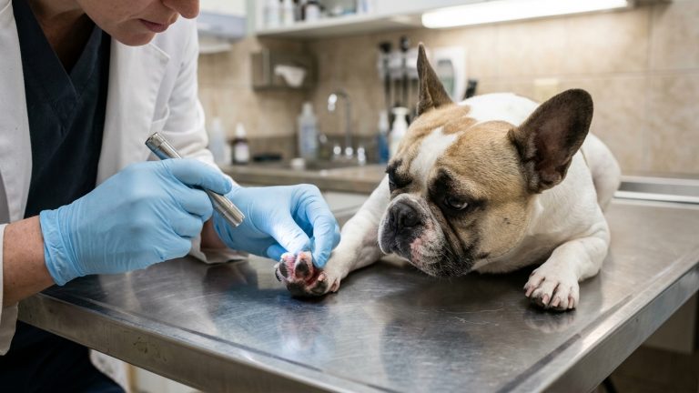
- The Withdrawal Reflex Trap (Spinal Reflex vs. Conscious Deep Pain): Owners and inexperienced examiners frequently pinch a paralyzed dog’s toe, see the leg pull back, and falsely conclude the dog “still feels its toes.” Pulling the leg back is merely a local L4–S1 spinal cord arc (the withdrawal reflex) that requires zero connection to the brain; true Deep Pain Perception requires a conscious cerebral response (turning the head, whining, dilating pupils, or attempting to bite) when hemostats compress the periosteum of the digit.
- Upper Motor Neuron (T3–L3) vs. Lower Motor Neuron (L4–S3) Bladder: Most thoracolumbar disc extrusions occur between T11 and L3, creating a spastic Upper Motor Neuron (UMN) bladder with high urethral sphincter tone that is difficult to express manually and overflows only when dangerously distended, whereas L4–S3 lesions produce a flaccid, dribbling Lower Motor Neuron bladder.
- Progressive Hemorrhagic Myelomalacia (10–15% of Grade 5 Dogs): In a subset of Grade 5 dogs, peracute vascular rupture triggers an ascending and descending self-propagating ischemic liquefaction of the spinal cord over 2 to 7 days—evidenced by cranial migration of the cutaneous trunci skin-twitch cutoff, loss of hindlimb withdrawal reflexes, abdominal wall flaccidity, and eventual phrenic nerve (C5–C7) respiratory failure.
- Fenestration of Adjacent Calcified Discs: During surgical hemilaminectomy, prophylactic blade or power fenestration of adjacent mineralized thoracolumbar discs (T11–L4) significantly reduces lifetime recurrence rates in Dachshunds and French Bulldogs.
Emergency Triage Matrix: Clinical Presentation & Required Response
| Triage Level | Clinical Signs & Presentation | Immediate Action Protocol |
|---|---|---|
| Tier 1: Immediate Neurosurgical Emergency (0–6 Hours) | Inability to stand or support weight on the hindlimbs (or all four limbs in cervical IVDD), dragging rear legs like a seal, loss of voluntary urination, or rapidly worsening hindlimb weakness over hours. | Confine immediately inside a rigid small carrier, withhold all food/water and oral medications (so anesthesia and imaging are not delayed), and transport directly to a 24-hour veterinary neurology center with MRI/CT and neurosurgery capability. |
| Tier 2: Urgent Same-Day Veterinary Evaluation (2–12 Hours) | Arched tense hunched back (kyphosis) or rigid low head carriage with neck spasms, crying when lifted, reluctance to jump or use stairs, or mild hindlimb wobbliness/paw knuckling while still able to walk (Grade 1–2). | Enforce immediate 100% crate rest (carry the dog outdoors on a short leash for bathroom only) and schedule a same-day veterinary neurological exam and multimodal analgesia plan (gabapentin, methocarbamol, and prescribed anti-inflammatory). |
| Tier 3: 4-to-6-Week Strict Crate Rest & Post-Op Rehabilitation | Grade 1–2 dog under conservative medical management OR post-hemilaminectomy Grade 3–5 dog recovering at home. | Maintain 24/7 confinement inside a 3×4-foot recovery crate or pen for the full 4 to 6 weeks (even after pain resolves at day 5, as the torn annulus fibrosus takes 6 weeks to scar), express the bladder every 6–8 hours if non-ambulatory, and perform guided PROM exercises. |
MRI vs. Plain Radiographs, The 6-Week Annulus Fibrosus Healing Rule & Bladder Expression
A crucial diagnostic reality for caregivers is that standard survey spinal radiographs (X-rays) cannot visualize the spinal cord or confirm which specific intervertebral disc has acutely extruded; calcified disc spaces visible on plain X-rays are frequently chronic, asymptomatic bystander discs, while the actual compressing disc material lies at an adjacent space. Definitive pre-operative localization requires cross-sectional advanced imaging—principally Magnetic Resonance Imaging (MRI, the gold standard that simultaneously reveals intramedullary T2-weighted hyperintensity and early myelomalacia) or Computed Tomography (CT)—performed immediately prior to surgical hemilaminectomy or ventral slot decompression under the same anesthetic episode.
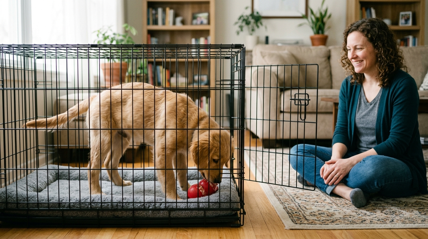
For Grade 1 and stable mild Grade 2 patients managed conservatively without surgery, the single most common cause of catastrophic relapse into Grade 4 or Grade 5 paralysis is ending crate rest prematurely. Once gabapentin, methocarbamol (for paraspinal muscle spasm), and a veterinary-prescribed NSAID relieve pain within 48 to 72 hours, the dog feels energetic and wants to run—yet the tear in the fibrous outer ring of the disc (annulus fibrosus) remains wide open. Any sudden twist, shake, or jump onto a couch during weeks 1 through 4 forces the remaining calcified nucleus pulposus straight through the unhealed rent into the spinal cord. Full fibrous scar formation across the annulus fibrosus requires a non-negotiable minimum of 4 to 6 weeks of strict crate confinement.
- Polyethylene Glycol & High-Dose Methylprednisolone Sodium Succinate (MPSS): Randomized clinical trials by the Canine Spinal Cord Injury Consortium (CANSORT-SCI) proved that high-dose intravenous methylprednisolone succinate (MPSS) offers zero neuroprotective benefit in Grade 5 dogs while causing severe gastrointestinal hemorrhage.
- Manual Bladder Expression Mechanics (Every 6–8 Hours): Any dog that cannot stand and walk independently (Grades 3–5) lacks voluntary micturition; caregivers must be trained in hands-on two-handed abdominal bladder palpation and gentle steady expression every 6 to 8 hours to prevent detrusor muscle overstretching (atony) and ascending urinary tract infections.
- Cervical IVDD (“Neck Disc”) Specifics: Extrusions at C2–C7 often cause excruciating root-signature forelimb pain and screaming spasms with minimal paresis (because the cervical vertebral canal is wide relative to cord diameter), or tetraparesis affecting all four limbs; neck collars must be permanently replaced with harnesses.
- Lifetime Furniture Ramp & Weight Mandate: Once an IVDD dog completes recovery, permanently install carpeted low-angle ramps at couches/beds, block open staircases with baby gates, and maintain a lean Body Condition Score (4/9) to reduce axial disc loading.
Strict Rule S1 Clinical Red Flags: Canine Thoracolumbar & Cervical Intervertebral Disc Disease (Hansen Type I & Type II IVDD)
Critical safety boundaries for Canine (Canis lupus familiaris — Chondrodystrophic Breeds: Dachshunds, French Bulldogs, Corgis & Beagles) caregivers:
- Zero Home NSAID or Aspirin Dosing Before the Vet Exam: Never give your dog a leftover carprofen, meloxicam, Galliprant, or baby aspirin tablet before heading to the emergency clinic; a single dose of aspirin or NSAID creates a dangerous drug-interaction conflict and can cause fatal gastric perforation under neurogenic spinal stress.
- Zero Chiropractic Spinal Manipulation on Acute IVDD Dogs: Never allow high-velocity chiropractic adjustments or vigorous back stretching on a dog with suspected intervertebral disc extrusion; manual spinal manipulation can drive unstable calcified disc fragments deeper into the spinal cord, causing instant irreversible paralysis.
Clinical Preparation & Biosecurity Action Checklist
- Place a suspected IVDD dog immediately inside a hard-sided travel carrier or laundry basket lined with a flat pad; lift the carrier smoothly while keeping the dog’s spine completely horizontal.
- Withhold food and water prior to emergency clinic arrival so advanced imaging (MRI/CT) and decompressive surgery can proceed immediately without aspiration risk.
- Prepare a ground-level 4-to-6-week recovery enclosure (a wire crate or small exercise pen no larger than 2×3 feet for a small dog) with non-slip yoga-mat flooring and water clipped to the bars.
- Ask the veterinary technician to demonstrate and supervise your hands-on manual bladder expression technique in the clinic before taking a non-ambulatory dog home.
Related Clinical Guides & Triage Protocols
Peer-Reviewed Clinical References & Evidence Grounding
- ACVIM consensus statement on diagnosis and management of acute canine thoracolumbar intervertebral disc extrusion (opens in new tab) (Journal of Veterinary Internal Medicine (2022) — ACVIM Consensus Statement by Olby, Moore, Brisson et al. indexed on NCBI PubMed (PMID: 36065965).)
- FGF4 retrogene on CFA12 is responsible for chondrodystrophy and intervertebral disc disease in dogs (opens in new tab) (Proceedings of the National Academy of Sciences / PNAS (2017) — Landmark genetic discovery by Brown, Dickinson, Bannasch et al. indexed on NCBI PubMed (PMID: 28990233).)
- American College of Veterinary Surgeons (ACVS): Intervertebral Disc Disease (IVDD) Grading & Surgical Hemilaminectomy (opens in new tab) (Board-certified veterinary surgical reference on Hansen Type I vs Type II pathology, deep pain nociception, and post-operative rehabilitation.)
- Merck Veterinary Manual: Intervertebral Disc Disease in Small Animals (opens in new tab) (Verified veterinary neurology reference on thoracolumbar and cervical disc extrusion localization, myelomalacia, and conservative vs surgical criteria.)
Source transparency
Reference mix for this guide
Source labels classify the institutional category of the references cited above.