This guide is general education for pet owners. If your pet has urgent symptoms, possible poisoning, injury, breathing trouble, collapse, severe pain, or sudden decline, contact a licensed veterinarian or local emergency service.
Editorial standard
How this guide was prepared
Dabbling waterfowl frequently ingest lead sinkers, galvanized wire, or metallic hardware that remains trapped in the muscular ventriculus under acidic mechanical grinding, causing delta-ALA dehydratase inhibition, intravascular hemolysis, and encephalopathic paresis.
Veterinary Clinical Notice (Domestic and Waterfowl Ducks (Anseriformes)): This peer-reviewed clinical reference addresses Avian Heavy Metal Toxicosis (Lead and Zinc) and Ventricular Hardware Disease and is grounded in verified veterinary literature. It does not substitute for hands-on physical examination, diagnostic imaging, or clinical pathology by a licensed exotic, avian, aquatic, or livestock veterinarian. Never attempt unguided invasive procedures or administer unprescribed pharmaceuticals when acute decompensation is present.
Clinical Quick Answer & Immediate Stabilization Protocol
Primary Clinical Directive for Avian Heavy Metal Toxicosis (Lead and Zinc) and Ventricular Hardware Disease: Patients in the Domestic and Waterfowl Ducks (Anseriformes) group mask early systemic illness until physiological reserves are exhausted. Immediate stabilization requires auditing enclosure thermal/environmental parameters, halting stressful handling or forced oral feeding in compromised animals, and arranging prompt veterinary diagnostics to address the underlying etiology before irreversible organ failure develops.
Pathophysiology, Etiology & Clinical Presentation in Domestic and Waterfowl Ducks (Anseriformes)
Clinical Evaluation of Waterfowl Heavy Metal Toxicosis and Ventricular Pathology
In veterinary toxicology—as outlined in references like the Merck Veterinary Manual—waterfowl (such as ducks, geese, and swans) are uniquely predisposed to heavy metal toxicosis, specifically lead (Pb) and zinc (Zn) poisoning.
Because of their foraging habits (dabbling or tipping up in aquatic sediments), waterfowl readily ingest spent lead shotgun pellets, discarded fishing sinkers, galvanized wire, coins, and other metallic debris. Once ingested, these objects pass into the muscular ventriculus (gizzard).
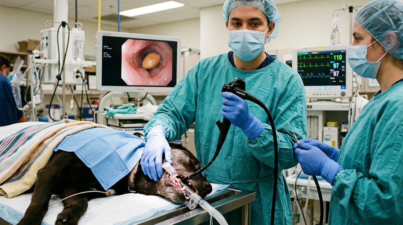
Pathophysiological Mechanism of the Ventriculus in Heavy Metal Poisoning
The ventriculus acts as the primary mechanical breakdown organ in avian digestion, utilizing powerful muscular contractions and ingested grit to grind food. Unfortunately, this physiologic process accelerates the destruction of ingested toxic metals:
- Erosion and Absorption: The high-pressure, acidic environment of the upper gastrointestinal tract combined with the grinding action of the gizzard rapidly breaks down and erodes metallic lead and zinc objects into soluble salts.
- Systemic Release: These bioavailable forms are then passed into the lower GI tract, absorbed into the bloodstream, and distributed to soft tissues (liver, kidneys, brain) and bone.
- “Hardware Disease” Parallels: While hardware disease classically refers to traumatic reticuloperitonitis in ruminants (where sharp metallic objects puncture the reticulum wall), avian species suffer a different kind of mechanical-chemical trauma. In birds, sharp or heavy foreign bodies trapped in the ventriculus can cause local mechanical ulcers, erosion of the koilin layer, secondary infections, and continuous, fatal systemic heavy-metal toxicosis.
Clinical Signs in Domestic and Waterfowl Ducks Patients
Avian heavy metal toxicosis typically manifests as a combination of gastrointestinal, neurologic, and hematologic dysfunction:
- Gastrointestinal: Anorexia, severe weight loss (“going light”), green-stained or watery diarrhea, regurgitation, and impacted or atonic ventriculus.
- Neurologic: Weakness, ataxia (impaired coordination), depression, drooping wings, tremors, paresis, and seizures.
- Hematologic: Lead specifically interferes with heme synthesis, frequently resulting in a nonregenerative, microcytic, hypochromic anemia.
Diagnosis in Domestic and Waterfowl Ducks Patients
- Radiography (Diagnostic Imaging):
- Whole-body or targeted abdominal radiographs are crucial. Because lead and zinc are radiopaque, orthogonal radiographs will readily reveal bright, metallic densities, fragments, or ingested grit outlines trapped within the silhouette of the ventriculus.
- Caveat: The absence of visible metal on a radiograph does not completely rule out toxicosis, as small fragments may have completely dissolved, or the bird may have passed the object after systemic absorption occurred.
- Blood/Tissue Metal Concentrations:
- Confirming elevated whole blood lead levels or tissue concentrations (liver/kidney analysis post-mortem) establishes a definitive diagnosis.
- Hematology & Biochemistry:
- Checking for evidence of anemia, basophilic stippling (in some species), and elevated AST, uric acid, or CK values.
Management and Treatment
According to standard veterinary toxicology protocols:
- Removal of Foreign Material: If metallic fragments are visualized in the ventriculus and the patient is stable enough, endoscopic retrieval, surgical ventriculotomy, or specialized flushing/dietary bulk (e.g., psyllium) may be utilized to encourage the passage or removal of residual metal.
- Chelation Therapy:
- Calcium Disodium Versenate (Ca EDTA): Frequently administered subcutaneously or intramuscularly (with adequate fluid therapy) to bind circulating lead and enhance its excretion via the kidneys.
- DMSA (Dimercaptosuccinic Acid) or D-Penicillamine: Often used for oral follow-up outpatient chelation therapy once acute stabilization is achieved.
- Supportive Care: Aggressive fluid therapy to protect renal function, nutritional support (gavage feeding if anorexic), and management of neurologic symptoms (e.g., diazepam or midazolam for seizures).
Emergency Triage Matrix: Clinical Presentation & Required Response
| Triage Level | Clinical Signs & Presentation | Immediate Action Protocol |
|---|---|---|
| Emergency | Seizures, inability to stand, green/watery diarrhea, severe weakness, head tremors. | Immediate veterinary critical care. Requires radiographs to confirm metal in the ventriculus and chelation therapy (CaEDTA). |
| Urgent | Lethargy, decreased appetite, mild incoordination or drooping wings. | Remove from free-range area to prevent further ingestion. Seek veterinary diagnostics within 12-24 hours. |
Differential Diagnosis, Clinicopathologic Workup & Husbandry Prevention
In avian and wildlife veterinary medicine, the management and pathophysiology of heavy metal toxicosis in Anseriformes (waterfowl such as ducks, geese, and swans) encompass critical diagnostic, clinical, and ecological challenges.
The interconnected concepts span toxicological comparison, pathology, intervention, diagnostics, and prevention:
Lead (Pb) vs. Zinc (Zn) Toxicosis in Waterfowl
Both heavy metals cause acute and chronic intoxication in Anseriformes, often resulting from the ingestion of foreign objects encountered during bottom-feeding. However, their sources, cellular targets, and clinical presentations differ:
- Lead Toxicosis (Plumbism):
- Source: Ingested spent lead shotgun pellets, lead fishing sinkers, or industrial paint/mining waste.
- Pathophysiology: Lead mimics calcium and binds to sulfhydryl groups in enzymes, severely disrupting heme synthesis (causing non-regenerative anemia), peripheral and central nervous systems, and renal function.
- Clinical Signs: Profound weakness, “dropped wing” posture, ataxia, green-stained watery feces, and severe emaciation (“reach-and-wasting syndrome”) due to muscular atrophy and anorexia.
- Zinc Toxicosis:
- Source: Ingested galvanized metals (wire, hardware, pennies minted after 1982, or cage components).
- Pathophysiology: Excess zinc damages the gastrointestinal tract, pancreas, and red blood cells, frequently resulting in severe hemolytic anemia and hemoglobinuria.
- Clinical Signs: Anorexia, lethargy, severe depression, diarrhea, polyuria, and acute hemolytic crisis. While both cause anemia, zinc heavily targets the pancreas and induces profound gastrointestinal ulceration/necrosis.
Proventricular Dilatation and Gastrointestinal Impairment
- Gastrointestinal Stasis: Waterfowl possess a powerful muscular gizzard (ventriculus) designed to grind hard items and grit. When toxic heavy metal particles (lead shot or zinc fragments) lodge in the gizzard or ventriculus, they are slowly ground down, accelerating systemic absorption.
- Proventricular/Ventriculus Impact: Chronic heavy metal toxicosis causes damage to the local enteric nervous system and smooth muscle walls. This leads to profound gastrointestinal hypomotility, stasis, and subsequent functional dilatation of the proventriculus and ventriculus. Food accumulates, causing regurgitation, foul-smelling fluid buildup, profound weight loss, and a palpable, impacted gizzard on physical examination.
Diagnostic Confirmation: Blood Lead Assay
- Sample Choice: Whole blood anticoagulated with EDTA or heparin is the definitive sample of choice for a blood lead assay. Serum or plasma are inappropriate because over 90% of circulating lead in birds is bound directly to erythrocytes (red blood cells).
- Interpretation:
- Background/Normal: <0.10 ppm (parts per million) or <10 µg/dL.
- Subclinical exposure: 0.20 to 0.49 ppm.
- Clinical Toxicity: >0.50 ppm, with severe, life-threatening or neurological toxicosis often exceeding 1.0 ppm.
- Adjunct diagnostics: Often paired with radiographs (to visualize radiopaque metallic densities in the ventriculus) and complete blood counts (CBC showing non-regenerative anemia with basophilic stippling and nucleated red blood cells).
Endoscopic Retrieval and Medical Management
When heavy metal fragments remain trapped in the upper GI tract (proventriculus or ventriculus) without having completely dissolved, prompt removal is vital to halt continuous systemic absorption.
- Endoscopic Retrieval: Rigid or flexible endoscopy allows direct visualization and retrieval of metallic fragments using specialized grasping forceps or baskets via the esophagus. This avoids invasive surgical ventriculotomy (gizzard surgery), which carries a higher risk of dehiscence and post-operative mortality in compromised waterfowl.
- Chelation Therapy: Endoscopy must be paired with medical chelation (e.g., Calcium Disodium EDTA (CaNa2EDTA) or succimer / DMSA) to bind circulating heavy metals in the bloodstream and enhance renal excretion.
- Supportive Care: Fluid therapy, nutritional support via gavage feeding (if safe), and vitamin supplementation (especially B-complex and Vitamin E/Selenium).
Wetland Foraging Safety and Ecosystem Health
Because Anseriformes are dabbling or diving bottom-feeders, they readily ingest substrate sediments, sifting through mud where spent lead ammunition and heavy metal pollutants accumulate over decades.
- Population Impacts: Lead poisoning causes high mortality and severe sub-lethal reproductive, behavioral, and immunological deficits across migratory waterfowl flyways. Carcasses are rapidly scavenged, meaning natural mortality events often go unnoticed by field managers.
- Mitigation & Safety: Transitioning completely from lead to nontoxic shot alternatives (such as bismuth, steel, tungsten-iron, or tin) for hunting, remediating contaminated wetland substrates, restricting lead fishing tackle use, and enforcing stringent environmental protection policies are essential steps to secure wetland foraging safety and safeguard global waterfowl populations.
Rapid-Response Clinical Red Flags: Avian Heavy Metal Toxicosis (Lead and Zinc) and Ventricular Hardware Disease
Critical stabilization boundaries for Domestic and Waterfowl Ducks (Anseriformes) caretakers:
- Zero Forced Oral Administration in Debilitated Patients: Never syringe-feed fluids, oils, or nutritional slurries to a dyspneic, obtunded, or hypothermic Domestic and Waterfowl Ducks (Anseriformes), as aspiration pneumonia and acute pharyngeal trauma carry high mortality.
- Thermal & Microclimate Stabilization Before Transit: Correct acute hypothermia or hyperthermia gradually during transport using insulated carriers and digital thermal monitoring calibrated specifically for Domestic and Waterfowl Ducks (Anseriformes).
Diagnostic Preparation & Husbandry Audit Checklist for Domestic and Waterfowl Ducks (Anseriformes)
- Record exact ambient, basking, or water temperature and humidity/water-quality metrics using calibrated digital probes prior to the veterinary appointment.
- Bring a fresh fecal sample, water sample, or photograph of the current enclosure layout, substrate depth, lighting fixtures, and diet labels for clinical evaluation.
- Document the precise timeline of anorexia, postural changes, respiratory effort, or abnormal excretions to assist differential staging.
- Prepare a dedicated, easily disinfected hospital or quarantine enclosure free of loose particulate hazards for post-visit recovery.
Related Clinical Guides & Triage Protocols
Peer-Reviewed Clinical References & Evidence Grounding
- Lead poisoning in wild waterfowl in Japan (opens in new tab) (Journal of wildlife diseases (1999) — Peer-reviewed clinical investigation indexed on NCBI PubMed (PMID: 10574537).)
- High Prevalences of Lead Poisoning in Wintering Waterfowl in Spain (opens in new tab) (Archives of environmental contamination and toxicology (1998) — Peer-reviewed clinical investigation indexed on NCBI PubMed (PMID: 9680527).)
- One Health: Children, Waterfowl, and Lead Exposure in Northwestern Nigeria (opens in new tab) (The Journal of the American Osteopathic Association (2017) — Peer-reviewed clinical investigation indexed on NCBI PubMed (PMID: 28556859).)
- Merck Veterinary Manual: Toxicoses Of Pet Birds (opens in new tab) (Verified veterinary reference retrieved via Antigravity Ground-Truth Search (merckvetmanual.com).)
Source transparency
Reference mix for this guide
Source labels classify the institutional category of the references cited above.