This guide is general education for pet owners. If your pet has urgent symptoms, possible poisoning, injury, breathing trouble, collapse, severe pain, or sudden decline, contact a licensed veterinarian or local emergency service.
Dosages must be calculated by a licensed veterinarian based on species, weight, and bloodwork. Never administer human medications or self-adjust prescription doses for pets.
Editorial standard
How this guide was prepared
Sudden lameness in dogs requires systematic evaluation to distinguish between minor soft tissue injuries and catastrophic orthopedic failure. A limping dog is exhibiting a clinical sign of pain or mechanical dysfunction. The diagnostic challenge involves determining whether the underlying pathology is a bone fracture, a muscular or ligamentous sprain, or an acute rupture of the cranial cruciate ligament.
Clinical Safety Notice: This protocol provides observational triage guidelines for canine lameness. It does not replace radiographic diagnostics or professional veterinary orthopedic assessment. Severe trauma involving non-weight-bearing lameness requires immediate professional intervention.
Quick Answer: Assessing Sudden Lameness
Canine lameness is classified by the degree of weight-bearing function and the presence of localized inflammation or instability. The primary distinction caregivers must observe is whether the dog is weight-bearing (even partially) or strictly non-weight-bearing. Non-weight-bearing lameness, especially following blunt trauma or accompanied by systemic signs such as pallor or lethargy, constitutes a Tier 1 emergency necessitating immediate stabilization and radiographic imaging.
Clinical Triage Matrix: Lameness Assessment
| Triage Tier | Clinical Presentation | Action Required |
|---|---|---|
| Tier 1: Immediate Emergency (0-2h) | Visible bone deformity or protruding bone (open fracture), non-weight-bearing with severe swelling after trauma, limping with pale gums or rapid breathing (internal injury), dragging limb with no pain response (nerve damage/spinal). | Proceed to emergency veterinary hospital immediately. Do not manipulate the limb. |
| Tier 2: Same-Day (12-24h) | Persistent non-weight-bearing lameness >24h without trauma, sudden hind leg lameness with “toe-touching” gait (cruciate rupture pattern), swollen hot joint with fever (septic arthritis), progressive lameness in growing large breed puppy (panosteitis/OCD). | Schedule urgent veterinary appointment. Restrict movement entirely. |
| Tier 3: Home Monitoring | Mild limping that improves with rest over 24-48h, slight favoring after vigorous exercise, intermittent skip in hind leg gait (luxating patella). | Strict cage rest and leash walking only. Monitor for 48 hours. |
Dangerous Home Pitfalls: What NOT to Do (Rule S1)
- NEVER give ibuprofen, aspirin, or acetaminophen: Administration of human non-steroidal anti-inflammatory drugs (NSAIDs) to dogs rapidly induces severe gastrointestinal ulceration, acute renal failure, and hepatotoxicity.
- NEVER splint a suspected fracture without training: Improper splint application worsens bone displacement, restricts vascular flow causing tissue necrosis, and dramatically increases patient pain.
- NEVER force exercise on a limping dog: Continuing activity on a compromised limb can convert a partial ligament tear into a complete rupture or displace an otherwise stable fracture.
- NEVER massage a hot swollen joint: Massaging a potentially septic (infected) joint or acute soft tissue injury exacerbates inflammation and can systemically spread infection or dislodge a local thrombus.
Differentiating Fracture, Sprain, and Cruciate Tear
The etiology of sudden lameness dictates the diagnostic approach and therapeutic intervention. Caregivers must observe the onset, duration, and mechanical presentation of the gait deficit.
Bone Fractures: Appendicular Trauma
Fractures typically result from significant kinetic energy transfer, such as vehicular trauma, high falls, or severe crush injuries. However, pathologic fractures can occur with minimal force if the underlying osseous structure is compromised by neoplasia (e.g., osteosarcoma) or metabolic disease. The hallmark of a complete fracture is acute, absolute non-weight-bearing lameness.
Clinical signs include pronounced focal swelling, palpable crepitus (a grinding sensation indicating bone-on-bone friction), visible angular deformity, and intense localized hyperesthesia (pain). If the fracture compromises the integument (an open fracture), the risk of osteomyelitis requires aggressive intravenous antimicrobial therapy and surgical debridement alongside stabilization. For further reading on managing severe trauma and bleeding, refer to our protocol on canine hemorrhage and wound first aid.
Soft Tissue Sprains and Strains
Sprains involve the stretching or tearing of ligaments (tissue connecting bone to bone), whereas strains affect muscles and tendons (tissue connecting muscle to bone). These injuries frequently follow athletic exertion, slipping on slick surfaces, or abrupt directional changes. Unlike fractures, mild to moderate sprains often present with partial weight-bearing lameness.
The patient may exhibit a shortened stride, reluctance to climb stairs, or stiffness after periods of recumbency. Palpation might reveal mild, diffuse edema rather than the acute, localized pain of a fracture. Initial management involves strict activity restriction. Diagnostics are primarily utilized to exclude osseous pathology, as soft tissues are poorly visualized on standard radiographs without advanced modalities like magnetic resonance imaging (MRI).
Cranial Cruciate Ligament (CCL) Tears
The canine cranial cruciate ligament is functionally analogous to the human anterior cruciate ligament (ACL). However, canine CCL disease is predominantly a degenerative biomechanical process rather than a purely acute athletic injury. It is a primary cause of hind limb lameness. When the ligament fails, the tibia displaces cranially relative to the femur during weight-bearing, causing profound mechanical instability and pain.
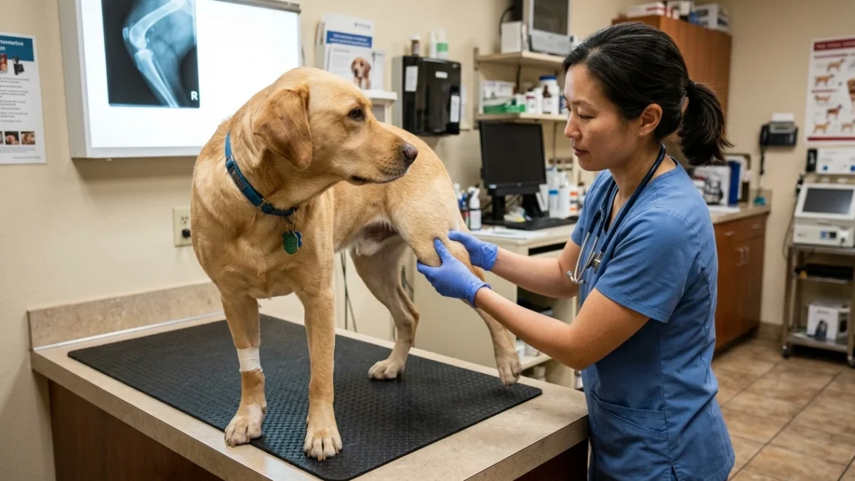
The classic presentation is an acute onset of hind limb lameness where the dog adopts a “toe-touching” posture—resting the tip of the paw on the ground without bearing significant load. A pathognomonic diagnostic maneuver performed by veterinarians is the evaluation of the “cranial drawer sign.” This involves manually assessing the abnormal forward translation of the tibia beneath the femur. Due to the precision required and the potential to induce pain, this test is exclusively a veterinary diagnostic tool and should not be attempted by caregivers.
The Role of Radiographic Evaluation
Diagnostic imaging is non-negotiable in the evaluation of severe or persistent lameness. While physical examination can isolate the origin of pain, radiographs (X-rays) provide critical objective data regarding osseous integrity, joint effusion, and degenerative changes.
X-rays are mandatory in cases of suspected fractures, chronic progressive lameness (to rule out primary bone tumors), or lameness unresponsive to conservative management. In the context of CCL disease, radiographs demonstrate secondary indicators such as joint effusion (loss of the infrapatellar fat pad shadow) and periarticular osteophytosis, confirming the diagnosis even if the ligament itself is radiolucent. Identifying osteoarthritis early is crucial; for ongoing management, see our guide on silent pain signs in senior dog arthritis.
Management and Intervention Protocols
Therapeutic strategy hinges precisely on the definitive diagnosis. Conservative management for sprains involves strict crate rest, controlled leash walks solely for elimination, and veterinary-prescribed canine-specific analgesia.
Fracture repair necessitates rigid stabilization. Depending on the fracture classification (e.g., transverse, comminuted, spiral), surgical intervention utilizing internal fixation (plates, screws, intramedullary pins) or external skeletal fixation may be required to achieve anatomical alignment and primary bone healing.
Surgical intervention is similarly the standard of care for CCL rupture, particularly in medium to large breed dogs. Techniques such as Tibial Plateau Leveling Osteotomy (TPLO) or Tibial Tuberosity Advancement (TTA) dynamically stabilize the stifle joint by altering biomechanical forces, rather than relying on static ligament replacement.
Frequently Asked Questions
What is the difference between non-weight-bearing lameness and a mild dog limp?
Non-weight-bearing lameness (carrying the leg completely off the ground without putting any weight on it) indicates severe structural pathology: a bone fracture, joint luxation, or complete rupture of the cranial cruciate ligament (CCL). A mild limp with partial weight-bearing may represent a minor sprain or early osteoarthritis.
Why is a sudden hind-leg limp often a cranial cruciate ligament (CCL) tear in dogs?
The cranial cruciate ligament stabilizes the canine stifle (knee). Unlike human ACL injuries caused by sudden trauma, canine CCL ruptures typically result from progressive ligament degeneration. When the weakened ligament snaps during routine activity, the tibia slips forward relative to the femur, producing sudden acute lameness and joint effusion.
Can I give my limping dog human aspirin, ibuprofen, or Tylenol for pain?
Never administer human pain relievers to a limping dog. Ibuprofen and naproxen cause acute stomach ulcers and kidney failure, while acetaminophen causes severe liver damage and methemoglobinemia. Always crate-rest your dog in a confined space and consult your veterinarian for safe, canine-specific prescription analgesics.
Sources and Clinical Evidence
This protocol relies on established veterinary orthopedic principles and peer-reviewed literature. The etiology of cruciate ligament disease and the necessity of appropriate imaging modalities are documented across current veterinary medical literature.
- PMID: 25146654 – Cranial cruciate ligament disease is a leading cause of hind limb lameness in dogs, often presenting as sudden non-weight-bearing lameness.
- PMID: 32675661 – Immediate radiographic evaluation is necessary for non-weight-bearing lameness following trauma to identify or rule out appendicular fractures.
- PMID: 28844883 – Administration of non-steroidal anti-inflammatory drugs intended for humans, such as ibuprofen, to dogs can result in severe gastrointestinal ulceration and acute renal failure.
Caregiver Safety Checklist: Lameness Management
- Confirm whether the dog is weight-bearing, partially weight-bearing, or non-weight-bearing.
- Examine the affected limb visually for angular deformity or protruding bone without manipulating it.
- Check mucous membrane color (gums) and respiratory rate to rule out systemic shock.
- Enforce immediate and strict rest; confine the dog to a small, secure area.
- Prevent access to stairs, furniture, and slippery flooring.
- Schedule immediate veterinary evaluation for any non-weight-bearing lameness or signs of trauma.
Final Safety Reminder: Lameness is a primary indicator of physiological compromise. Avoid delaying professional diagnostics, and strictly adhere to the contraindication against administering human pharmacological agents to canine patients.
Source transparency
Reference mix for this guide
Source labels describe the type of organization behind each reference; they are not a claim that any outside organization reviewed this article.
- canine hemorrhage and wound first aid Reference source · newspet.net
- silent pain signs in senior dog arthritis Reference source · newspet.net
- Dog Shaking & Lethargic Differential Triage Reference source · newspet.net
- PMID: 25146654 Government or public agency · pubmed.ncbi.nlm.nih.gov
- PMID: 32675661 Government or public agency · pubmed.ncbi.nlm.nih.gov
- PMID: 28844883 Government or public agency · pubmed.ncbi.nlm.nih.gov