This guide is general education for pet owners. If your pet has urgent symptoms, possible poisoning, injury, breathing trouble, collapse, severe pain, or sudden decline, contact a licensed veterinarian or local emergency service.
Dosages must be calculated by a licensed veterinarian based on species, weight, and bloodwork. Never administer human medications or self-adjust prescription doses for pets.
Editorial standard
How this guide was prepared
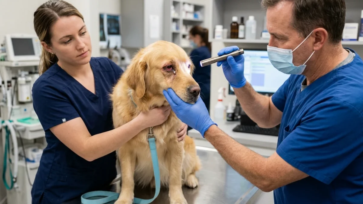
A red, squinting eye in a dog represents a time-sensitive clinical presentation requiring immediate differentiation between self-limiting conjunctivitis and sight-threatening conditions like acute glaucoma or deep corneal ulcers. Rapid elevation in intraocular pressure causes irreversible blindness within hours, making prompt veterinary evaluation the primary mandate for sudden unilateral ocular distress.
Quick Answer: Differentiating Canine Red Eye
Blepharospasm (squinting), epiphora (excessive tearing), and episcleral injection (redness) represent non-specific signs of severe ocular pain. Acute glaucoma typically presents with a fixed, dilated pupil and a globe that feels firm upon palpation. Corneal ulcers exhibit extreme light sensitivity and rapid progression if infected. Uncomplicated conjunctivitis generally spares vision, features normal pupil size, and presents primarily with diffuse redness and discharge. Treat any acute squinting as a high-level emergency until intraocular pressure is verified as normal.
Clinical Triage Matrix: Canine Ocular Distress
| Triage Tier | Clinical Presentation | Action Required |
|---|---|---|
| Tier 1: Immediate Emergency (0-2h) | Acute glaucoma (rock-hard eye, dilated fixed pupil, vision loss within hours), penetrating eye injury, chemical splash, sudden complete eye closure with visible corneal defect. | Proceed immediately to veterinary ER. Transport with minimal stress. Do not apply pressure to the face. |
| Tier 2: Same-Day Evaluation (12-24h) | Red eye with mucopurulent discharge lasting longer than 48 hours, squinting with visible white spot on cornea (ulcer), cherry eye (prolapsed third eyelid gland). | Schedule primary care visit today. Use Elizabethan collar to prevent self-trauma. |
| Tier 3: Home Monitoring | Mild watery discharge with no squinting, slight redness after windy walk, occasional pawing at eye with normal vision. | Observe for 24 hours. Flush gently with sterile canine saline. Seek care if signs progress. |
Pathophysiology of the Canine Red Eye
Ocular redness results from the engorgement of conjunctival or episcleral vessels. Recognizing the exact source of this vascular congestion guides the clinical index of suspicion. Deep episcleral injection signals intraocular disease, whereas superficial conjunctival hyperemia points toward surface irritation.
Acute Canine Glaucoma: The Silent Blinder
Primary glaucoma occurs when the iridocorneal angle fails to drain aqueous humor effectively, precipitating a catastrophic rise in intraocular pressure (IOP). Normal canine IOP ranges between fifteen and twenty-five millimeters of mercury. Glaucomatous crises push these readings above fifty millimeters of mercury. This extreme pressure rapidly ischemicizes the optic nerve and retina. Clinical signs manifest abruptly. Owners report sudden lethargy, anorexia, and intense eye pain. The affected eye appears enlarged (buphthalmos) with a cloudy, bluish cornea (corneal edema). The pupil remains fixed and dilated, non-responsive to light stimulation. Differentiating this state from generalized systemic collapse requires careful observation; review dog shaking and lethargic differential triage for broader context.
Corneal Ulceration: Surface Defects and Infection Risk
The cornea is densely innervated, rendering any epithelial defect excruciatingly painful. Ulcers arise from trauma, distichiasis (abnormal eyelashes), keratoconjunctivitis sicca (dry eye), or underlying metabolic conditions. Superficial abrasions cause intense squinting and tearing. If opportunistic bacteria invade the stroma, melting ulcers develop. These aggressive infections dissolve collagen, progressing to spontaneous globe rupture within days. Diagnosis mandates fluorescein staining, which highlights the exposed hydrophilic stroma.
Canine Conjunctivitis: Inflammation of the Mucosa
True isolated conjunctivitis is frequently overdiagnosed. Most canine conjunctival inflammation occurs secondary to other primary defects, such as dry eye or environmental allergens. Presentation includes diffuse redness, varying amounts of mucoid or purulent discharge, and mild discomfort. Unlike felines, primary infectious conjunctivitis is relatively uncommon in canines, though viral and bacterial etiologies exist. Systemic allergic reactions also present with ocular signs, similar to the facial swelling seen in dog bee or wasp sting anaphylaxis.
Dangerous Home Pitfalls: What NOT to Do (Rule S1)
- NEVER use human eye drops containing tetrahydrozoline (Visine) or other vasoconstrictors on dogs.
- NEVER delay glaucoma evaluation by more than 24 hours; permanent blindness occurs rapidly.
- NEVER attempt to remove embedded foreign objects from the globe with tweezers or fingers.
- NEVER apply pressure to a possibly ruptured globe or forcefully open swollen eyelids.
- NEVER reuse old prescribed drops, especially those containing corticosteroids, without verifying the absence of a corneal ulcer.
Veterinary Diagnostics and Intervention Protocols
Arrival at a clinical facility triggers a systematic ophthalmic examination. The standard database includes Schirmer tear testing to quantify aqueous production, tonometry to measure intraocular pressure, and fluorescein staining to detect epithelial breaches.
When glaucoma is confirmed, immediate pressure reduction therapies commence. Intravenous osmotic diuretics shrink the vitreous volume. Topical therapies target aqueous production and outflow. If medical management fails to lower pressure, surgical interventions, including laser cyclophotocoagulation or enucleation, alleviate intractable pain.
Corneal ulcers receive targeted topical antibiotic therapy. Severe, deep ulcers necessitate surgical grafting using conjunctival flaps or biomaterials to provide structural support and a blood supply for healing. The frequency of topical application for aggressive ulcers exceeds once every two hours.
Sources and Evidence Basis
This clinical review integrates findings from peer-reviewed veterinary ophthalmology literature.
- PubMed: Diagnosis and management of canine glaucoma
- PubMed: Canine superficial corneal ulcers: clinical presentation and healing time
- PubMed: A review of canine conjunctivitis
Caregiver Safety Checklist: Immediate Actions
- Place an Elizabethan collar (cone) on the dog immediately to prevent rubbing.
- Confine the dog to a dimly lit, quiet room to reduce photophobia and stress.
- Do not wipe the eye with dry tissues; use sterile saline on a soft cotton pad if debris removal is necessary.
- Call the nearest emergency veterinary facility to confirm their ability to measure intraocular pressure.
- Prepare for transport, keeping the dog’s head elevated and avoiding pressure on the neck.
Early identification of ocular discomfort ensures the highest probability of preserving vision and anatomical integrity. Owners must remain vigilant for subtle signs of eye pain, including increased sleeping, altered behavior, or reluctance to allow facial contact. Differentiating benign irritation from surgical emergencies remains the exclusive domain of veterinary professionals equipped with diagnostic instrumentation.
Continued monitoring following any ocular incident dictates visual outcomes. Even seemingly minor abrasions deteriorate into vision-threatening ulcers if opportunistic pathogens colonize the defect. Adherence to prescribed therapy schedules—especially regarding the frequency of topical drop administration—dictates the pace and success of corneal healing.
Systemic conditions routinely manifest ocular signs. Hypertension, coagulopathies, and systemic infectious diseases alter the appearance of the anterior chamber, retina, or conjunctiva. A thorough physical examination accompanies the localized ophthalmic assessment. Recognizing the eye as an extension of the vascular and nervous systems reinforces the requirement for comprehensive diagnostic evaluation when facing unexplained ocular changes.
Protective strategies mitigate the incidence of traumatic eye injuries. Securing dogs during vehicular transport, avoiding dense brush during walks, and restricting aggressive play reduce the likelihood of penetrating trauma or chemical exposure. Proactive management of known breed-specific predispositions, such as routine intraocular pressure monitoring in susceptible terriers or spaniels, offers the best defense against catastrophic glaucomatous vision loss.
Source transparency
Reference mix for this guide
Source labels describe the type of organization behind each reference; they are not a claim that any outside organization reviewed this article.
- dog shaking and lethargic differential triage Reference source · newspet.net
- dog bee or wasp sting anaphylaxis Reference source · newspet.net
- PubMed: Diagnosis and management of canine glaucoma Government or public agency · pubmed.ncbi.nlm.nih.gov
- PubMed: Canine superficial corneal ulcers: clinical presentation and healing time Government or public agency · pubmed.ncbi.nlm.nih.gov
- PubMed: A review of canine conjunctivitis Government or public agency · pubmed.ncbi.nlm.nih.gov