This guide is general education for pet owners. If your pet has urgent symptoms, possible poisoning, injury, breathing trouble, collapse, severe pain, or sudden decline, contact a licensed veterinarian or local emergency service.
Dosages must be calculated by a licensed veterinarian based on species, weight, and bloodwork. Never administer human medications or self-adjust prescription doses for pets.
Editorial standard
How this guide was prepared
Oviductal dystocia in laying hens occurs when an egg stalls within the shell gland (uterus) or vagina due to ionized hypocalcemia, salpingitis, or malformed shells, compressing pelvic nerves, obstructing cloacal excretion, and risking fatal pressure necrosis.
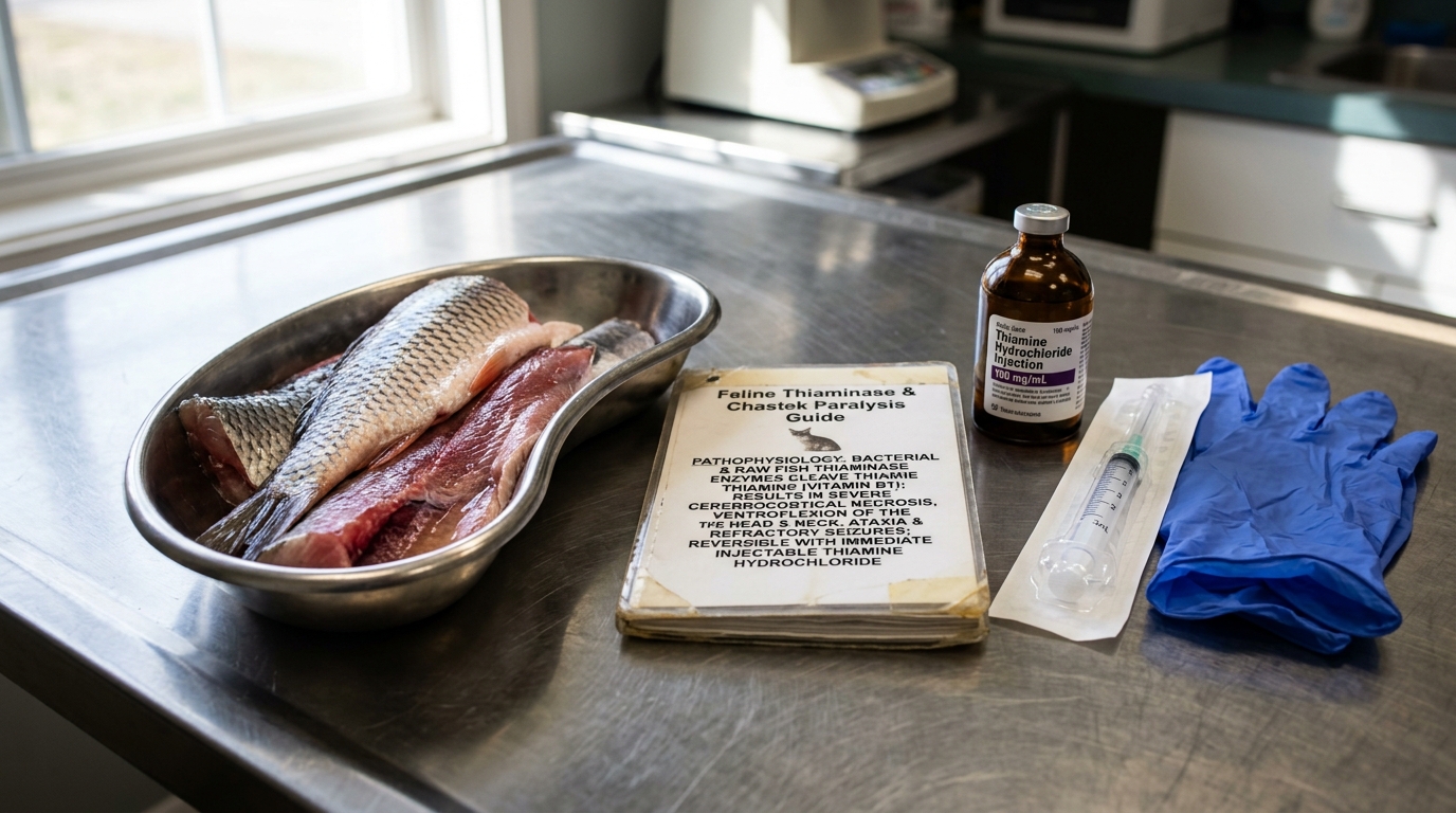
Veterinary Clinical Notice (Domestic Chicken (Gallus gallus domesticus)): This peer-reviewed clinical reference addresses Avian Oviductal Dystocia (Egg Binding) and Coelomic Egg Yolk Peritonitis and is grounded in verified veterinary literature. It does not substitute for hands-on physical examination, diagnostic imaging, or clinical pathology by a licensed exotic, avian, aquatic, or livestock veterinarian. Never attempt unguided invasive procedures or administer unprescribed pharmaceuticals when acute decompensation is present.
Clinical Quick Answer & Immediate Stabilization Protocol
Primary Clinical Directive for Avian Oviductal Dystocia (Egg Binding) and Coelomic Egg Yolk Peritonitis: Patients in the Domestic Chicken (Gallus gallus domesticus) group mask early systemic illness until physiological reserves are exhausted. Immediate stabilization requires auditing enclosure thermal/environmental parameters, halting stressful handling or forced oral feeding in compromised animals, and arranging prompt veterinary diagnostics to address the underlying etiology before irreversible organ failure develops.
Pathophysiology, Etiology & Clinical Presentation in Domestic Chicken (Gallus gallus domesticus)
In backyard chickens (Gallus domesticus), reproductive disorders such as egg binding, oviductal dystocia, hypocalcemia, and coelomic peritonitis are closely linked, often acting as cascading events. Veterinary literature (such as the Merck Veterinary Manual and peer-reviewed studies indexed on PubMed) outlines the pathophysiology, diagnostics, and integrated treatment protocols for these emergencies.
Pathophysiology and Interrelationships
- Egg Binding & Oviductal Dystocia: Egg binding is the failure of a fully formed egg to pass through the oviduct (shell gland/uterus or vagina) at a normal rate. Dystocia represents a more advanced state involving physical or mechanical obstruction. Causes in chickens include oversized or double-yolked eggs, misshapen eggs, soft-shelled eggs, obesity, lack of exercise, or structural fatigue of the oviductal smooth muscle.
- Hypocalcemia: Calcium is required for effective myometrial (uterine) contractions during oviposition. Inadequate dietary calcium, lack of vitamin D3/UV exposure, or high egg-laying exhaustion leads to hypocalcemia or calcium tetany. Without adequate ionized calcium, the smooth muscle of the oviduct cannot contract with enough force to expel the egg.
- Coelomic Peritonitis (Egg Yolk Peritonitis): If an egg ruptures inside the oviduct or coelomic (abdominal) cavity, or if reverse peristalsis forces yolk material and bacteria (most commonly Escherichia coli) backward out of the oviduct, severe serosal inflammation ensues. Yolk acts as an exceptional growth medium for bacteria, rapidly escalating a sterile chemical peritonitis into a septic, life-threatening coelomic infection characterized by fibrin accumulation and caseous, cooked-yolk exudate.
Clinical Presentation in Domestic Chicken Patients
Affected backyard hens typically present with:
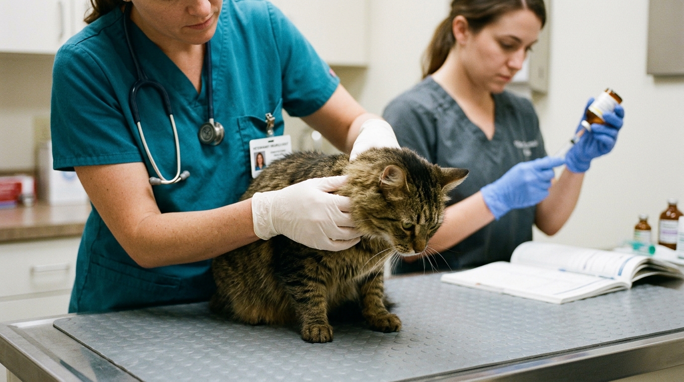
- Depression, lethargy, fluffed feathers, and sitting at the bottom of the coop or nest box.
- An upright “penguin-like” vertical stance due to abdominal discomfort or weight distribution.
- Straining, pumping of the tail, abdominal distension, and respiratory distress (dyspnea) resulting from compression of the air sacs by a large or impacted mass.
- Posterior paresis or paralysis in advanced cases due to the egg pressing against the lumbosacral plexus or secondary hypocalcemic tetany.
- Vent/cloacal prolapse if prolonged, futile straining occurs.
Diagnostic Imaging: Radiography
Radiography is a cornerstone diagnostic modality:
- Dorsoventral (DV) and Lateral Projections: A standard “bird-in-box” or tabletop radiograph will easily identify radiopaque, calcified mineralized shells within the coelomic cavity.
- Findings: Radiographs can confirm the presence, size, number, and exact anatomical location of retained eggs. They also help differentiate true egg binding from other causes of coelomic distension, such as ascites, internal laying (pure coelomic peritonitis without a shell), organomegaly, or coelomic neoplasms. Soft-shelled or unmineralized eggs may require contrast studies or ultrasonography, as they are radiolucent.
Comprehensive Medical and Surgical Treatment Protocols
According to standard avian and poultry veterinary guidelines:
A. Stabilization First (Supportive Care)
Before attempting to move or force an egg out, the patient must be stabilized:
- Fluid Therapy: Correct dehydration via parenteral (subcutaneous or intravenous) balanced electrolyte solutions (e.g., Lactated Ringer’s Solution).
- Environment: Provide a warm, humid environment (approx. 85–90°F / 29–32°C with high humidity) to relax smooth muscle and reduce shock.
- Supplemental Calcium: Administer parenteral calcium gluconate (typically veterinary-calibrated weight-specific dosing IM or slow IV/IO, diluted to avoid cardiac arrhythmias) regardless of immediate blood ionized calcium levels, to restore oviductal contractility.
B. Medical Management & Assisted Delivery
- Analgesia & Anti-inflammatories: Non-steroidal anti-inflammatory drugs (NSAIDs) like meloxicam help manage pain and reduce edema in the cloaca and pelvic canal.
- Lubrication: Gentle infusion of sterile water-soluble lubricants or liquid paraffin directly into the cloaca/vagina can ease passage.
- Warm Soaks: Warm water baths (hydrotherapy) targeting the lower abdomen can relax the vent sphincter muscles.
- Medical Stimulation: Oxytocin or arginine vasotocin and prostaglandins (e.g., Prostaglandin E2 gel) are sometimes used only after hypocalcemia is corrected and if there is absolute confirmation that the oviduct is unobstructed, as forcing contractions against a mechanical blockage can cause uterine rupture.
C. Minimally Invasive & Surgical Options
- Manual Manipulation: Under sedation or general anesthesia, gentle, steady caudoventral digital pressure can sometimes ease the egg through the vent.
- Ovocentesis (Aspiration): If the egg cannot be passed intact and presents a continuous obstruction, percloacal ovocentesis can be performed. A needle (e.g., 18-gauge) attached to a syringe is carefully introduced through the vent and shell to aspirate the yolk and albumen, allowing the shell to be collapsed and gently extracted in fragments without tearing the mucosal lining.
- Surgery (Salpingohysterectomy / Coelomotomy): Reserved as a last resort for cases involving severe oviductal torsion, ectopic eggs, unresolving coelomic peritonitis, or eggs firmly adhered/ruptured within the salpinx. Concurrent treatment for coelomic peritonitis (systemic broad-spectrum antimicrobials, coelomic lavage if surgical) is mandatory if yolk or bacterial contamination has leaked into the body cavity.
Emergency Triage Matrix: Clinical Presentation & Required Response
| Triage Level | Clinical Signs & Presentation | Immediate Action Protocol |
|---|---|---|
| Emergency | Straining to push with no result, wide ‘penguin’ stance, pale comb, lethargy, prolapsed tissue. | Immediate veterinary intervention for calcium gluconate injection and safe manual extraction. Do not crush the egg. |
| Urgent | Sitting in the nesting box for 12+ hours, reduced appetite, tail pumping. | Provide a warm Epsom salt bath (15-20 minutes) and place in a dark, quiet, humid isolation kennel. |
Differential Diagnosis, Clinicopathologic Workup & Husbandry Prevention
Avian reproductive medicine encompasses the oviductal physiology, dystocia pathology, surgical intervention, and flock management practices of the domestic hen (Gallus domesticus).
Normal Physiological Background
- Anatomy & Egg Formation: A healthy laying hen possesses a functional left ovary and oviduct. The oviduct is divided into the infundibulum (site of fertilization), magnum (albumen/egg white deposition), isthmus (shell membranes), and the shell gland (uterus).
- Calcium Carbonate Layer: In the shell gland, the developing egg is encased in a hard shell composed primarily of calcium carbonate (CaCO3). This process requires massive mobilization of calcium from both the diet and specialized medullary bone reserves.
- Ration & Nutrition: A proper layer ration must balance energy, protein, and minerals (specifically a high ratio of coarse calcium and adequate Vitamin D3) to support continuous shell mineralization and prevent metabolic fatigue.
- Lighting Management: Photoperiod (day length) and light intensity directly stimulate the hypothalamic-pituitary-gonadal axis, controlling hormone release (estrogen and luteinizing hormone) that triggers ovulation. Improper lighting management—such as increasing day length too rapidly in pullets—can force birds into premature production before their skeletal and reproductive tracts are fully mature.
Pathology & Clinical Complications
When nutrition, lighting, or management fails, or when physical/genetic hurdles arise, a cascade of reproductive disorders can occur:
- Egg Binding (Dystocia): This occurs when a fully formed egg fails to pass through the oviduct or cloaca at a normal rate.
- Primary drivers: Hypocalcemia (calcium deficiency leads to weak uterine muscle contractions and soft-shelled eggs), obesity (narrowing the pelvic canal), oversized or double-yolked eggs, and premature photostimulation in young pullets.
- Oviductal Prolapse: Continuous, severe straining from egg binding or muscular exhaustion can cause the lower oviduct and cloacal tissues to evert and protrude outside the vent. Exposed tissues are rapidly contaminated, leading to edema, necrosis, and pecking by flockmates.
- Egg Yolk Peritonitis: This condition arises when yolk material or fully formed eggs are released into the abdominal coelom (abdominal/internal laying) rather than being properly captured by the infundibulum, or when an impacted oviduct ruptures. The presence of yolk in the sterile peritoneal cavity incites severe, sterile or secondary bacterial inflammation (peritonitis), fluid accumulation (ascites), systemic toxemia, and a characteristic “penguin-like” upright posture.
Medical and Surgical Treatment
- Conservative Care: Mild cases of egg binding can sometimes be managed with warm baths, local lubrication, calcium gluconate supplementation (to stimulate uterine contraction), and manual gentle manipulation by an experienced handler or veterinarian. Prolapse cases require cleaning, reduction of the swelling (using hyperosmolar solutions like sugar water or ice packs), and temporary retention sutures (purse-string).
- Salpingohysterectomy: In advanced, recurrent, or severe surgical emergencies—such as chronic egg yolk peritonitis, severe oviduct rupture, uterine tumors, or irreproducible prolapse/impaction—a salpingohysterectomy (surgical removal of the oviduct and uterus) may be performed by an avian veterinarian to save the bird’s life.
Prevention via Flock Management
Preventing these cascading reproductive failures relies heavily on foundational husbandry:
- Lighting Protocols: Strictly control photoperiods during the rearing phase. Avoid increasing day length prematurely for growing pullets to ensure adequate body weight, pelvic development, and skeletal calcium stores are achieved prior to the onset of lay.
- Nutritional Balance: Provide a balanced layer ration containing precise calcium-to-phosphorus ratios. Supplementing free-choice oyster shell ensures hens have adequate calcium carbonate available overnight when shell calcification peaks.
- Weight Control: Prevent flock obesity through proper feed restriction or energy dilution, maintaining proper pelvic elasticity and reducing the incidence of dystocia.
Rapid-Response Clinical Red Flags: Avian Oviductal Dystocia (Egg Binding) and Coelomic Egg Yolk Peritonitis
Critical stabilization boundaries for Domestic Chicken (Gallus gallus domesticus) caretakers:
- Zero Forced Oral Administration in Debilitated Patients: Never syringe-feed fluids, oils, or nutritional slurries to a dyspneic, obtunded, or hypothermic Domestic Chicken (Gallus gallus domesticus), as aspiration pneumonia and acute pharyngeal trauma carry high mortality.
- Thermal & Microclimate Stabilization Before Transit: Correct acute hypothermia or hyperthermia gradually during transport using insulated carriers and digital thermal monitoring calibrated specifically for Domestic Chicken (Gallus gallus domesticus).
Diagnostic Preparation & Husbandry Audit Checklist for Domestic Chicken (Gallus gallus domesticus)
- Record exact ambient, basking, or water temperature and humidity/water-quality metrics using calibrated digital probes prior to the veterinary appointment.
- Bring a fresh fecal sample, water sample, or photograph of the current enclosure layout, substrate depth, lighting fixtures, and diet labels for clinical evaluation.
- Document the precise timeline of anorexia, postural changes, respiratory effort, or abnormal excretions to assist differential staging.
- Prepare a dedicated, easily disinfected hospital or quarantine enclosure free of loose particulate hazards for post-visit recovery.
Related Clinical Guides & Triage Protocols
Peer-Reviewed Clinical References & Evidence Grounding
- Percloacal Ovocentesis in the Treatment of Avian Egg Binding: Review of 20 Cases (opens in new tab) (Journal of avian medicine and surgery (2019) — Peer-reviewed clinical investigation indexed on NCBI PubMed (PMID: 31893620).)
- Reproductive Disorders in Parrots (opens in new tab) (The veterinary clinics of North America. Exotic animal practice (2017) — Peer-reviewed clinical investigation indexed on NCBI PubMed (PMID: 28169184).)
- Resolution of egg binding is possible in most client-owned parrots when multiple treatment strategies are considered (opens in new tab) (Journal of the American Veterinary Medical Association (2025) — Peer-reviewed clinical investigation indexed on NCBI PubMed (PMID: 39938214).)
- Merck Veterinary Manual: Egg Bound Or Impacted Oviducts In Poultry (opens in new tab) (Verified veterinary reference retrieved via Antigravity Ground-Truth Search (merckvetmanual.com).)
Source transparency
Reference mix for this guide
Source labels classify the institutional category of the references cited above.