This guide is general education for pet owners. If your pet has urgent symptoms, possible poisoning, injury, breathing trouble, collapse, severe pain, or sudden decline, contact a licensed veterinarian or local emergency service.
Editorial standard
How this guide was prepared
Fossorial horned frogs absorb water and solutes directly across their vascularized ventral pelvic patch; accumulated nitrogenous waste or chemical residues in stagnant substrate trigger acute transcutaneous neurotoxicity, erratic hindlimb spasms, andanasarca.
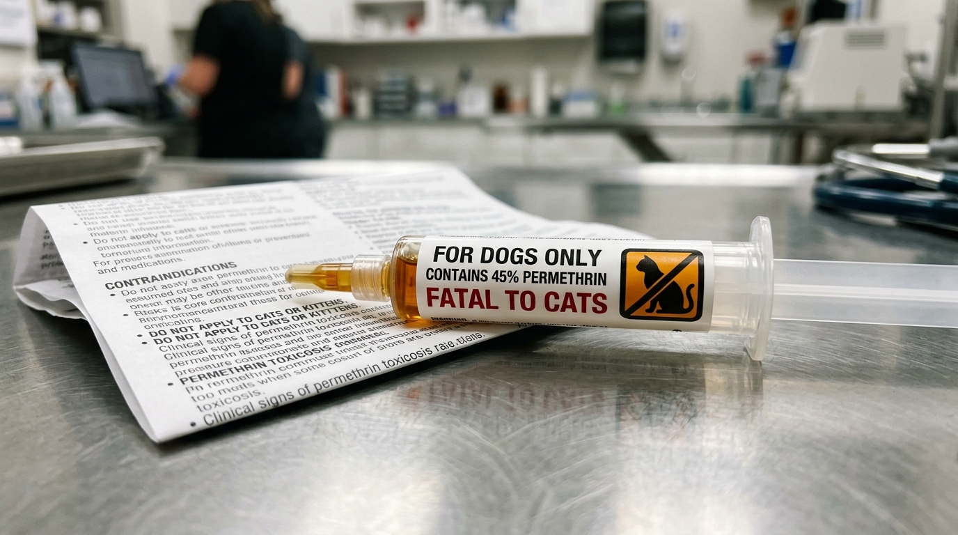
Veterinary Clinical Notice (South American Horned Frog (Ceratophrys cranwelli & C. ornata)): This peer-reviewed clinical reference addresses Anuran Autointoxication Syndrome (Toxic-Out Syndrome) and is grounded in verified veterinary literature. It does not substitute for hands-on physical examination, diagnostic imaging, or clinical pathology by a licensed exotic, avian, aquatic, or livestock veterinarian. Never attempt unguided invasive procedures or administer unprescribed pharmaceuticals when acute decompensation is present.
Clinical Quick Answer & Immediate Stabilization Protocol
Primary Clinical Directive for Anuran Autointoxication Syndrome (Toxic-Out Syndrome): Patients in the South American Horned Frog (Ceratophrys cranwelli & C. ornata) group mask early systemic illness until physiological reserves are exhausted. Immediate stabilization requires auditing enclosure thermal/environmental parameters, halting stressful handling or forced oral feeding in compromised animals, and arranging prompt veterinary diagnostics to address the underlying etiology before irreversible organ failure develops.
Pathophysiology, Etiology & Clinical Presentation in South American Horned Frog (Ceratophrys cranwelli & C. ornata)
“Toxic-out syndrome” (frequently discussed in amphibian husbandry and exotic veterinary medicine regarding Ceratophrys species, commonly known as Pacman frogs or horned frogs) describes a severe, acute form of autointoxication.
Pathophysiology: How It Happens
- Transcutaneous Absorption and Excretion: Amphibian skin is highly permeable, acting as a respiratory and fluid-exchange membrane. This unique physiology means frogs both absorb substances from their environment and excrete nitrogenous waste (primarily urea and ammonia) directly through their skin.
- Substrate Fouling & Ammonia Build-up: If a frog is kept in an enclosure with poorly maintained, unwashed, or overly-saturated substrate (soil, coconut fiber, or moss) that accumulates high levels of urine, feces, and decomposing organic matter, the concentration of environmental toxins—especially ammonia—spikes.
- Reversal of Concentration Gradient: Normally, waste moves outward from the frog’s body. When external ammonia and waste concentrations in the fouled substrate become excessively high, the osmotic and chemical gradient reverses or stalls.
- Autointoxication: The frog begins absorbing environmental ammonia and accumulated toxins backward through its skin (transcutaneous absorption) faster than it can process or clear them.
Clinical Signs (The Toxidrome)
When systemic levels of ammonia and waste overwhelm the frog’s nervous and osmoregulatory systems, it triggers a distinct constellation of signs often referred to by keepers and vets:
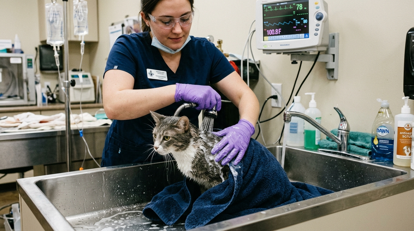
- Erratic Kicking / Spastic Limb Extensions: Sudden, violent, or uncoordinated kicking and twitching of the hind legs, often triggered by neurological irritation and pain/muscle cramping.
- Edema / Bloating: Profound fluid retention and generalized swelling as the frog’s lymphatic and renal systems fail to regulate fluid balance.
- Lethargy alternating with Hyperactivity: Periods of profound weakness/apathy broken up by sudden, erratic jumping or spasms.
- Discoloration or Cloudy Eyes: Corneas may appear cloudy or milky, and the skin may look overly slimy, irritated, or stripped of its protective mucus coat.
Management and First Aid
While comprehensive diagnostic criteria for exotic species are outlined in resources like the Merck Veterinary Manual (under amphibian environmental toxicities and husbandry-related diseases), immediate field and veterinary treatment generally prioritizes halting further absorption and promoting elimination:
- Immediate Decontamination (Clean Water Soaks): Transfer the frog immediately to a shallow bath of clean, unchlorinated, dechlorinated water to encourage passive elimination and dilute toxins across the skin.
- Frequent Water Changes: The soak water must be replaced regularly (every 20 to 60 minutes initially) because the frog will quickly leach toxins back into the surrounding shallow water.
- Total Environmental Reset: Completely discard and replace the contaminated enclosure substrate. Thoroughly sanitize the enclosure before returning the animal to a clean, properly monitored environment.
- Veterinary Care: If neurological signs (spasms, severe rolling, failure to right itself) persist, a veterinarian experienced in reptile and amphibian medicine should be consulted for supportive therapies, fluid therapy, or management of secondary electrolyte imbalances.
Emergency Triage Matrix: Clinical Presentation & Required Response
| Triage Level | Clinical Signs & Presentation | Immediate Action Protocol |
|---|---|---|
| Emergency | Violent erratic jumping, stretching hind legs rigidly, severe bloating, unresponsiveness. | Immediate transfer to a bath of pure, dechlorinated spring water. Requires emergency veterinary fluids to flush toxins. |
| Urgent | Restless behavior, unusually pale or dark skin, refusing food. | Completely replace the substrate immediately. Soak the frog in shallow, clean, dechlorinated water. |
Differential Diagnosis, Clinicopathologic Workup & Husbandry Prevention
In clinical amphibian medicine for Ceratophrys species (ornate or Argentine horned frogs/Pacman frogs), “toxic-out” syndrome (autointoxication), hydrocoelom, renal and lymph heart failure, and emergency remediation strategies such as dechlorinated spring water flushes, activated carbon filtration, and strict substrate replacement schedules govern environmental toxicology and osmoregulatory stabilization.
“Toxic-Out” vs. Autointoxication
- The Concept: In herpetoculture, “toxing out” (or colloquial autointoxication) refers to a systemic syndrome where an amphibian is poisoned by high concentrations of environmental toxins—most frequently accumulated nitrogenous waste (ammonia, urea), decomposing organic matter, or chemical residues absorbed directly through its highly permeable skin and specialized pelvic patch.
- Mechanism: Amphibians do not drink water in the traditional sense; they absorb it osmotic-dermally along with whatever solutes are present in their substrate or pooling water. When captive substrates become foul, stagnant, or overloaded with urates and feces, the chemical gradient reverses or overwhelms the frog’s capacity to excrete toxins, leading to neurotoxic and osmoregulatory collapse (manifesting as hyperactivity followed by lethargy, spastic hind-leg extension, skin sloughing, and systemic shock).
Hydrocoelom, Renal Failure, and Lymph Heart Failure
- Hydrocoelom: This is the abnormal accumulation of fluid within the coelomic (abdominal) cavity. In horned frogs, it is often visibly apparent as severe, symmetric abdominal distension (“water belly”).
- The Physiological Cascade:
- Renal & Lymphatic Integration: Unlike mammals, amphibians rely heavily on specialized pulsatile organs called lymph hearts (pulsatile muscular structures near the urostyle and spine) to pump interstitial fluid and lymph back into the venous circulation, working in tandem with the kidneys to manage fluid and electrolyte homeostasis.
- The Pathophysiology: When an environmental toxin (“toxic-out”), systemic infection, or primary renal disease damages kidney function or depresses lymph heart output, the delicate fluid balance fails. Lymphatic drainage grinds to a halt, and fluid leaks massively into interstitial spaces (lymphedema) or pools inside the body cavity (hydrocoelom). This mimics a form of congestive or circulatory failure unique to amphibians, where hydrostatic and oncotic pressures completely destabilize.
Therapeutic and Environmental Interventions
When managing or preventing these linked crises, targeted environmental remediation is required:
- Spring Water Flush:
- Role: Mineral-balanced, chlorine/chloramine-free spring water (or properly remineralized RO water) is used to perform immediate, repeated static baths or partial tank resets.
- Application: Acute “toxed-out” frogs are often pulled from contaminated enclosures and placed into shallow, clean spring-water quarantine setups (water level reaching no higher than mid-thorax to prevent drowning if the frog is weak). Frequent water changes flush out residual toxins absorbed via transdermal routes.
- Activated Carbon:
- Role: In filtration systems or aquatic staging setups, activated carbon adsorbs dissolved organic compounds, tannins, medications, and chemical impurities.
- Application: While mostly utilized in semi-aquatic or water-feature setups, it ensures that recirculating water systems remain stripped of heavy chemical loads that could compromise an already fragile amphibian dermis.
- Substrate Replacement Schedule:
- The Prevention Protocol: Because Ceratophrys frogs are heavy-bodied, spend much of their time burrowing, and regularly release large loads of urea/waste directly into their soil, substrate fouling is the leading trigger for environmental autointoxication.
- Schedule Rules:
- Spot cleaning: Daily removal of urates, feces, and soiled substrate.
- Full substrate replacement: Depending on enclosure size, humidity, and drainage, organic bioactive or coco-coir/soil substrates in strict-captive setups generally require complete replacement every 4 to 6 weeks (or sooner if an ammonia/foul sulfur smell develops) to intercept toxic accumulation before a “toxic-out” or subsequent hydrocoelom event can manifest.
Rapid-Response Clinical Red Flags: Anuran Autointoxication Syndrome (Toxic-Out Syndrome)
Critical stabilization boundaries for South American Horned Frog (Ceratophrys cranwelli & C. ornata) caretakers:
- Zero Forced Oral Administration in Debilitated Patients: Never syringe-feed fluids, oils, or nutritional slurries to a dyspneic, obtunded, or hypothermic South American Horned Frog (Ceratophrys cranwelli & C. ornata), as aspiration pneumonia and acute pharyngeal trauma carry high mortality.
- Thermal & Microclimate Stabilization Before Transit: Correct acute hypothermia or hyperthermia gradually during transport using insulated carriers and digital thermal monitoring calibrated specifically for South American Horned Frog (Ceratophrys cranwelli & C. ornata).
Diagnostic Preparation & Husbandry Audit Checklist for South American Horned Frog (Ceratophrys cranwelli & C. ornata)
- Record exact ambient, basking, or water temperature and humidity/water-quality metrics using calibrated digital probes prior to the veterinary appointment.
- Bring a fresh fecal sample, water sample, or photograph of the current enclosure layout, substrate depth, lighting fixtures, and diet labels for clinical evaluation.
- Document the precise timeline of anorexia, postural changes, respiratory effort, or abnormal excretions to assist differential staging.
- Prepare a dedicated, easily disinfected hospital or quarantine enclosure free of loose particulate hazards for post-visit recovery.
Related Clinical Guides & Triage Protocols
Peer-Reviewed Clinical References & Evidence Grounding
- NCBI PubMed (PMID: 35422258) — The Amphibian Heart (Vet Clin North Am Exot Anim Pract, 2022) (opens in new tab) (Peer-reviewed veterinary clinical study indexed in NCBI PubMed (Vet Clin North Am Exot Anim Pract, 2022).)
- NCBI PubMed (PMID: 36965879) — Amphibian Dermatology (Vet Clin North Am Exot Anim Pract, 2023) (opens in new tab) (Peer-reviewed veterinary clinical study indexed in NCBI PubMed (Vet Clin North Am Exot Anim Pract, 2023).)
- NCBI PubMed (PMID: 31759449) — Amphibian Renal Disease (Vet Clin North Am Exot Anim Pract, 2020) (opens in new tab) (Peer-reviewed veterinary clinical study indexed in NCBI PubMed (Vet Clin North Am Exot Anim Pract, 2020).)
- Merck Veterinary Manual: Environmental Diseases Of Aquatic Animals In Aquatic Systems (opens in new tab) (Institutional veterinary reference and clinical practice guideline.)
Source transparency
Reference mix for this guide
Source labels classify the institutional category of the references cited above.